A Moment of Creativity: Cartoon cells in real-life

Very frequently I find myself staring at an object and seeing something else, something that may only be understood by me because of my background and past experiences. In such events it is frustrating to try and explain to others why I find that object so exciting. But sometimes I manage to get the message through.

While enjoying a morning in my apartment I came across something I thought was pretty cool. It reminded me of the French-Japanese animated television series Once Upon a Time… Life produced by Procidis (those unfamiliar with the program, click here). I eagerly followed every episode of this show as a kid, and did it again when the rerun was on. Looking back, I think it is incredible how much information can be squeezed into an educational TV show for kids (the same can be said another popular TV series from the same studio – Once Upon a Time… Man).
Anyway, back to my story – what I saw was a real-life representation of the way cells were depicted in the program. Without thinking too much, I grabbed my camera and tried to photograph what I have just seen.

caladium1

Cartoon cells. Or are they?


The similarity to the cells illustrated in the cartoon series is astonishing to me. The “cells” were actually flowers from one of my Caladium bicolor plants:

caladium2

Unopened male flowers of Caladium bicolor


To make the flowers red, I fired a flash through one of the semi-transparent leaves, which added some color.

caladium

Caladium bicolor flower and leaf

Petrophila: Salticid-mimic moths

A few months ago, I returned to Belize to conduct a small arachnid survey. While I was there, I took part in designing insect light traps for Caves Branch Jungle Lodge. The concept of a light trap is simple – flying nocturnal insects use the moonlight to navigate at night, and when there is a brighter light source present (like a light bulb) they are attracted to it. We wanted to have a few traps set up before the beginning of BugShot, and we ran a few trials in several locations using different lighting settings to see what works best and which insects show up at the traps. Very soon we realized that the traps attracted an impressive diversity of insects, but also their predators – spiders, frogs and bats quickly learned the locations of the traps and came regularly to feed. In several occasions fire ants showed up to raid the unsuspecting insects.

Petrophila sp. in typical resting posture, partially exposing the hindwings

Petrophila sp. in typical resting posture, partially exposing the hindwings


One of the insects that we saw in great numbers every night was a small, plain-looking moth from the family Crambidae. I would probably not pay attention to it if it were not for four black dots arranged in a row on the margin of each of its hindwings. Many moths rest with their hindwings concealed by the forewings, however these moths, belonging to genus Petrophila, had a unique body posture at rest, exposing only the dotted part of their hindwings. This pattern looked very familiar to me, but I could not pinpoint from where exactly. Then a few nights later one of these moths decided to rest pointing sideways with its head rather than upwards like most moths. And it finally hit me: this moth has an image of a jumping spider on its wings looking straight at you. The mimicry is so convincing that the moth wings even have hair-like scales where supposedly the spider’s head is.

Side view of Petrophila sp.

Side view of Petrophila sp.


I should be careful here. Pareidolia is a known phenomenon in which one searches for known patterns just about anywhere. It is what makes people see the face of Jesus Christ on a burnt piece of toast, it is what makes you see a face on a rocky terrain on Mars, and it what makes you see a number when looking at the wings of Diaethria species.
What I mean to say is that the color pattern on the wings of Petrophila species reminds me of a salticid spider, and perhaps it works the same for other animals as well. There is also a behavioral display that makes the mimicry even more deceiving: the moth moves its wings to mimic the movements of a jumping spider. In search for a second opinion, I turned to someone who breathes and sleeps jumping spiders. Thomas Shahan, who fortunately was around for BugShot, confirmed my suspicion and even came up with an ID for a possible model spider: a female Thiodina sp. And so we went on to find a jumping spider that looked like the one shown on the moths’ wings. In any case, to my untrained eyes it seems that this pattern is common in several moth genera, and in other insects as well. Some will debate whether this apparent image actually evolved to depict what we want it to be, but I can only imagine the reaction of a jumping spider to this image and behavior by the moth. Jumping spiders are known to have good vision; a jumping spider will stall to examine an opponent to avoid conflict. This may give the moth a few seconds to escape. A good analysis of similar mimicry in other species is discussed here.

A different species of Petrophila, recorded from the same light trap. This one is smaller and seems to have a slightly different spider image.

A different species of Petrophila, recorded from the same light trap. This one is smaller and seems to have a slightly different spider image.


The same Petrophila species as above, here with a possible salticid model - female Thiodina sp. from the same location in Belize. What I find amazing is that the wings even show some of the iridescence seen in the spider's eyes.

The same Petrophila species as above, here with a possible salticid model – female Thiodina sp. from the same location in Belize. What I find amazing is that the wings even show some of the iridescence seen in the spider’s eyes.


Petrophila moths are unique among Lepidoptera for having aquatic caterpillars. They occupy running freshwater habitats, rivers and streams, where they feed on algae by scraping the surface of submerged rocks and stones. The genus has a wide distribution across the Americas and many species occur in temperate zones in addition to tropical regions.

You know the moths are successful in their mimicry when you find others deploying the same strategy: Nectopsyche is a genus of caddisflies (order Trichoptera) that shows a similar pattern – moth-like adults have four tiny black spots arranged in a row at the margin of their forewings, along with pale stripes.

You know the moths are successful in their mimicry when you find others deploying the same strategy: Nectopsyche is a genus of caddisflies (order Trichoptera) that shows a similar pattern – moth-like adults have four tiny black spots arranged in a row at the margin of their forewings, along with pale stripes.


Not only moths, but also many other insects orders were represented in our trap catch. I hope that Caves Branch continues to make good use of these sturdy light traps to record the insects surrounding the lodge. There is great potential for scientific work to be done here.

Light trap in Caves Branch, Belize

Light trap in Caves Branch, Belize


My NZ ordeal (part 2)

Some time ago I wrote about my NZ accident and I mentioned that the story did not end there. One of the most frustrating experiences I had upon leaving NZ was a slow and thorough inspection of everything I had in my luggage by the customs officers. I am not sure what they were expecting to find, because I had collecting and import permits for all the research material I obtained. After discussing this with other visitors to NZ (not necessarily scientists) I learned that it is a standard procedure that some lucky individuals must endure. But imagine spending a couple of hours in an isolated part of the airport with other “suspects”, where you are being treated like trash for doing nothing. I will not go into details but it was definitely some of the most nerve-racking time I had in my life.

It was not before I returned to Canada when things started to take a wrong turn. I purposely delayed writing a post about it, mainly because I needed time to digest what has happened and to understand the details of my case. My plan was to write it down eventually because I believe it can be important for other graduate students facing a similar situation, and I think I am ready to share.

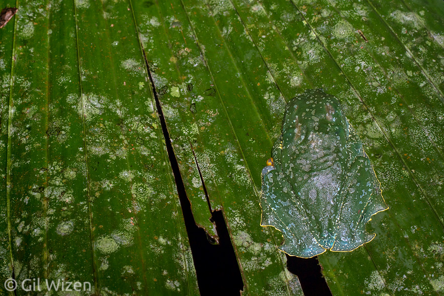
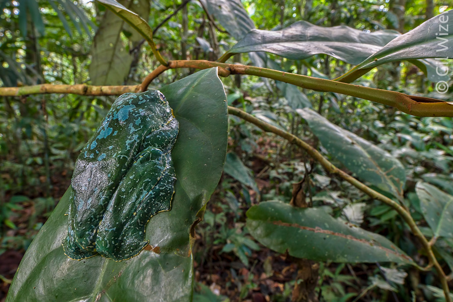
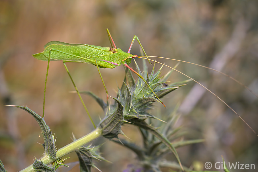
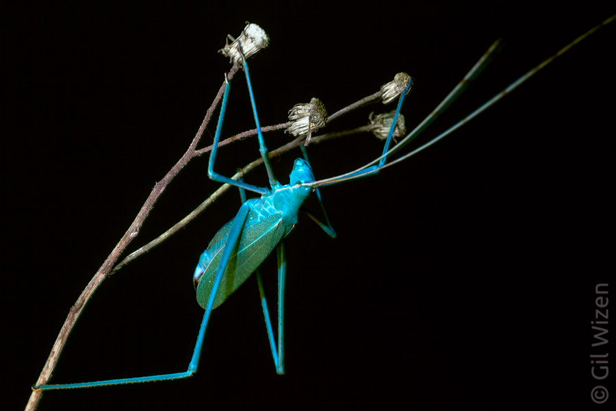
So cut back to early 2013, I spent several months in NZ, most of the time observing mating behavior of ground weta (ensiferan insects of the genus Hemiandrus) as a part of my PhD research, but I also found the time for experimenting with my photography. There is something about being all alone, in a foreign place, that sparks your creativity to try new and interesting ideas. Some of the shots I managed to capture in NZ were surprising even for me (see some of them here).

(Feel free to skip this paragraph if you only want to read the “juicy” parts of the story. It explains the research I was conducting in NZ)
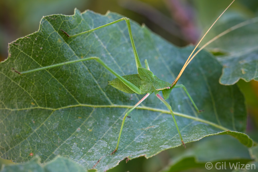
Before I detail my story, let me elaborate a bit on weta mating behavior. One of the things I aimed to capture was the mating process in “short-tailed” Hemiandrus species. “Short-tailed” means that, unlike most members of suborder Ensifera, the females do not possess a long ovipositor (a device used to inject the eggs into different substrates, such as soil, wood, leaves, etc’). This character was found to be associated with a high level of maternal care: the ground weta females seal themselves in an underground burrow, spending several months tending their eggs and the hatching nymphs. As for the mating process, in most ensiferan insects the male transfers a nuptial gift for the female to feed on during mating. This gift comes in the form of a protein-rich spermatophore, attached by the male to the female’s genitalia. In “short-tailed” ground weta however, the males deposit their nuptial gift on a modified segment on the females’ abdomen, and in some species they even display mate-guarding behavior while the females consume their nutritious gift.
The result of my photography trials was a series of shots that I am very proud of, showing the whole mating process:

Hemiandrus-mating1

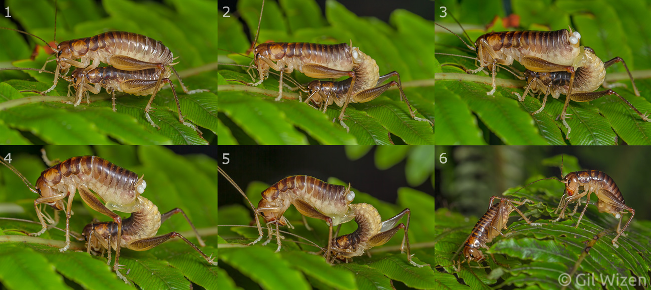
The mating process in Hemiandrus pallitarsis. 1. The male (bottom) attached to the female; 2. The male attaches the sperm ampullae to the female’s genitalia; 3. The male disconnects from the female’s genitalia and extends two phalli; 4. The male’s phalli start secreting the nuptial gift; 5. The nuptial gift is deposited on the female’s modified sixth abdominal segment; 6. The male displays mate-guarding behavior while the female consumes the nuptial gift.

It is important to mention that during my time in NZ this information about the ground weta mating behavior was already known and published. My intention was to use these photos in presentations and perhaps in my PhD thesis as a communication aid. And indeed, I presented them to several faculty members during a meeting and they were impressed.

From that point on things started to go downhill.
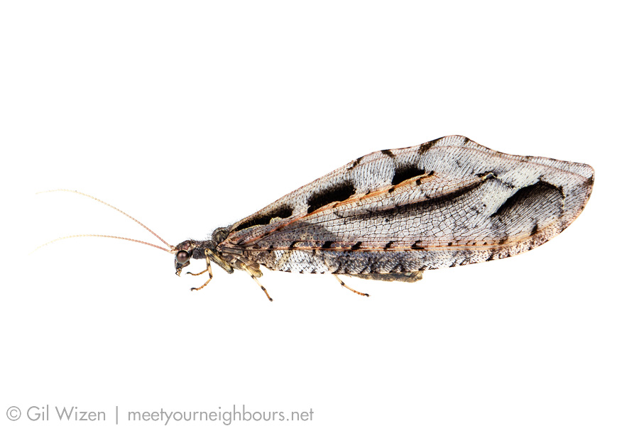
My PhD supervisor back then requested to use a photograph of a wasp for a textbook chapter he was working on, and I replied that I would gladly license it for publication. That is, after payment of a small licensing fee. Then happened the thing I was worried about the most: he asked how this sits with use of my weta photos in his future publications. My reply was the same.
I have always allowed the use of my photos for presentation purposes, whether it was an in-class presentation, conference talk, poster, etc’. My only issue is with publication and distribution of my photographs. This is a legal matter (Copyright Law is a real thing) that involves a license in order to manage who has copyrights over the use of the photo by transferring all or part of the copyrights from the photo owner to someone else. Anyone who is not familiar with this and those who wish to know more, you can refer to my Image Use page.

My refusal to give the photos away triggered an unfortunate chain of events that ended with the supervisor kicking me out of the lab and terminating his supervision, essentially shutting down my PhD research. I was accused of being greedy:


“Even after agreeing that I have been more than generous with funding all your New Zealand doctoral research and that your work and all expenses were, in fact, fully covered by my NSERC Discovery grant, you insisted on going ahead in charging me for the use of the photographs. It is for this reason alone that I no longer wish to supervise your doctoral research. While I think that you have the skills, background and experience necessary for tackling this project, I cannot continue to supervise a student with such a mercenary approach to the student-supervisor relationship…”


(As a side note I should say that this person now avoids mentioning this small detail and tells a different story, trying to make it look like my departure from his lab was a mutual decision. It was not. This infuriates me because I did want to go on with my PhD research. To him I say – take responsibility for your actions!)

I think the supervisor was missing a crucial detail of what copyright protection is meant for. Notice how nothing is mentioned about how much I was going to charge for the photos? That is because this was never discussed, the supervisor did not even bother to inquire about my image use policy, for him it was enough that I intended to charge for the use. Protecting my copyrights?? Nonsense, in his eyes I was all in it for the money! While there are photographers out there who routinely take copyright infringements to court in order to collect the damages, I cannot brag for having such a history.

What is even more surprising was that I was handed a copy of the university’s Intellectual Property Policy with a friendly remark that everything I create during my term as a graduate student is owned by the university. Really? Everything?…
Well, this is not exactly how it works. According to this policy, the university owns any idea, invention or pretty much any data that you collect or create while conducting research (raw data cannot be copyrighted anyway). This applies to any form of media that may contain such relevant data, including photos. But it is important to understand that while the university legally owns the data concealed within a photo (or a disk, flash drive, laptop etc’), it does not own the media itself unless it was the university’s property in the first place. In my case, the photos were captured by me using my photography gear, therefore the university did not own the photos and had no copyrights over use of the photos themselves. When requested, I provided low-resolution files for data acquisition purposes; nevertheless the university cannot use those photos in future publications without my consent.

Some will say that I should have agreed to give away the photos to maintain a healthy working relationship with my supervisor. This may sound like the right thing to do, however in my opinion a relationship in which someone is using you for their own personal gain is not a healthy relationship. Do take the time to think about it. Moreover, everyone has the right to choose whether they want to share their creation with someone else. I chose not to, and while my choice may seem strange to some people, it comes after a history of bad experiences. I learned my lesson the hard way, and I will not devalue my work any longer.

Here I turn to every grad student out there – you DO NOT owe anything to your supervisor other than working on your project. A supervisor cannot force you to give away your rights on something you created. If they do, that’s academic bullying. For example, if you produce an artwork piece depicting your research subject, does the university or your supervisor immediately own it? Of course not.
Universities as institutions have committees or unions that can advise grad students how to deal with such disputes. I know now that I should have taken this case straight to both the university’s Research Ethics Board and Copyrights Office. The end result of me moving to a different academic department might have been the same, but the supervisor’s disgraceful behavior would have been recorded on file, which could later act as a warning sign for prospective students.
Lastly, I think it is a real shame that a professor who spends so much time and energy fighting the disease of academic plagiarism is completely unaware of Copyright Law.

As for those ground weta photos, they will probably never see the light of day. I hope at least that you enjoyed viewing them here, and that you learned something about politics in academia at the same time.

Killing in the name of

About two weeks ago, Piotr Naskrecki, whose blog The Smaller Majority I routinely follow (and you should too), posted a nice story about his encounter with the world’s biggest and heaviest living spider, the South American Goliath Birdeater (Theraphosa blondi), while surveying a rainforest in Guyana. The blogpost gained respectable attention from everyone who appreciates a good natural history piece, but only when picked up by a “viral-content-spreading” website it started getting the full public attention it deserved. Or did it? To be honest, I do not know which website was first in line to spread the story of a “puppy-sized spider with foot-long legs” but it wasn’t long before the internet and the media snatched the story and made it spread like wildfire. The result was interesting but also horrifying to watch – almost within a day the internet was flooded with various reports and interpretations of the original blogpost, some of which were poorly written and included embarrassing inaccuracies. The story quickly climbed up in popularity and a few days ago was ranked #14 in the fastest spreading online news, along with other “popular” news stories such as the Ebola outbreak. Needless to say, the majority of those reports shamelessly used Piotr’s photographs on their own websites without permission.

A small insert about content turning viral: One simply cannot predict what will become viral on the internet. I have tried to do this myself and failed, when photographs that I thought were decent received no attention at all, whereas crappy photos that I took out of laziness just before I went to bed were instantly favored and shared. Want an example? Here are two:

Mother amblypygid (Paraphrynus raptator) protecting her babies

Mother amblypygid (Paraphrynus raptator) protecting her babies


Small-scaled Godzilla - baby ambush bug (Phymata monstrosa)

Small-scaled Godzilla – baby ambush bug (Phymata monstrosa)


When comments started pouring in on the tarantula article, the usual mix of positive (“amazing animal!”) and negative (“kill it with fire!”) responses could be seen. But among those there was a strong stream of comments calling for justice, as it was revealed that the spider was eventually collected for research and deposited in a museum collection. At first I did not know where this information originated from, after all the original post by Piotr did not include any statement about collecting the spider. Later that day I found it, in the closing paragraph of this report.

Not a South American Goliath Birdeater, but close enough; an adorable Ecuadorian Pinktoe Tarantula (Avicularia huriana)

Not a South American Goliath Birdeater, but close enough; an adorable Ecuadorian Pinktoe Tarantula (Avicularia huriana)


Spiders are sweet, I agree. This bashing response, however, points to an alarming problem. First, I do believe these comments truly come from people who care about nature and the environment. So why am I writing this? Because I think it is unclear to the public what scientists actually do, and in the case of biologists, why they collect data and specimens in the field and what happens to such specimens further along the road. The funny thing is that there are many blogs out there, run by scientists, trying to take a public outreach approach by explaining the routine and difficulties scientists face in their daily work. Among these blogs there are quite a few that discuss the topic of collecting insect specimens for research, like Biodiversity in Focus and Beetles in the Bush to name a few. However, I do not see people submitting the same type of preaching comments (promoting the insects’ rights to live) in these blogs. The reason is quite depressing: the general public, the same people who were exposed to the Goliath Birdeater story via the various viral news websites, do not read blogs about scientific research, even though these blogs are there for the public in the first place. Here is where Piotr’s blog is doing so well; it brings easily digestible information about the wonders of earth, in a language that can be understood by any person, without excessive technical details or jargon. The same can be said about his books. In addition, everyone loves a good photograph, and Piotr’s photos are nothing short of stunning.

So why bash a scientist for killing a single spider for research?

Piotr gave an excellent response to this issue in his subsequent post (now integrated within the original Goliath Birdeater post), I really could not have said it better myself, so make sure you head over to his blog to read it. I will just add a few things. For start, I do not think the accusing commenters are aware of Piotr’s significant contributions to nature conservation. Unfortunately, the finger is fast on the trigger keyboard, and it has become extremely easy to criticize any person one does not agree with on the internet. But the problem is much worse than trolling. Most people do not realize that the only reason they know what they know about nature, whether it is related to animals, plants or their environment, is because some scientist spent a lot of time in remote areas collecting this information, and then took the liberty of publishing it for the greater good. Without scientific knowledge no one would even know the spider in Piotr’s post is a Goliath Birdeater, it would just pass as a legendary giant arachnid. The only way to properly identify a species or describe a new one is to collect it and compare it to related species that were… also collected and killed previously. You see, from a scientific point of view, this work will never end. There are so many species out there, with many of them undescribed or unknown. Be thankful and considerate towards those who sacrifice so much of themselves not only to deliver these majestic creatures all the way to your computer screen at the comfort of your home or office, but also invest enormously towards protection of their natural habitat from destruction.

Insect art: rubber stamps

Some months ago I stumbled upon on a blog post in Japanese with many images of insect plush toys and wooden figurines. The topic was the annual Mozo Mozo exhibition in Japan. In case you have never heard about this exhibition, dedicated to the love of small creatures, every year various artists join to display and sell insect-themed artwork. The diversity of works ranges from drawings and figurines to toys, clothing, bags and accessories. I hope one day I have a chance to attend this event in person. Until then, I will have to settle for photos posted online.
Nevertheless, upon noticing some artwork that I really liked in those photos, I decided to try and contact several artists in hopes they still have something available that can be sent overseas.

This is how I discovered the amazing hand-made stamps by the talented Mayu Watanabe (check out her work here). This young artist is not only very capable of translating complex structures (not just insects) into rubber stamps, but she also finds original and intriguing ways to combine different stamping methods to create esthetically pleasing designs on almost any paper media, for example postcards or gift wrappings.

stamps2

Desert locust (Schistocerca gregaria), solitary form (left) and gregarious form (right). Beautiful stamp artwork by Mayu Watanabe


stamps1


stamps3

I couldn’t think of a better packing for these! Excellent!


The artist also puts a lot of thinking into the packaging of her artwork (marketing departments, take notes!): Every stamp is packed separately on cotton, stapled within a piece of cardboard and cellophane. Entomologists out there, does this sound familiar? This is exactly how you would send unmounted insect specimens overseas! I even had a prepared horned dung beetle (Copris sp.) lying around and compared it to its stamp counterpart – the similarity is very entertaining.

stamps4
But if you think this ends here, think again. Flip the packaged stamp and you will find the collecting data for that “specimen”, along with the species’ ID. Again, written very accurately according to the rules of scientific collecting.

stamps5
Overall, I am very impressed with the quality of these stamps, and I look forward to seeing more work from Mayu Watanabe in the future. I find it refreshing to see someone who not only loves what they are doing, but also does it very professionally.

Cruziohyla – a dream come true

In 2003 I visited Costa Rica as a part of my first trip to Latin America. One of the hostels I stayed at had a large poster hung featuring many Costa Rican frog species, to show the high amphibian diversity that is found in this beautiful country. This was the first time I saw a photo of a splendid leaf frog, Cruziohyla calcarifer. Back then it was called Agalychnis calcarifer but in 2005, following a revision in the Hylidae family, it was placed within a new genus, Cruziohyla, along with another species. Then in 2018, another revision to the genus was published, splitting Cruziohyla calcarifer into two species. The costa Rican species is now called Sylvia’s tree frog, Cruziohyla sylviae.
When I saw the photo I was stunned. It looked like a massive tree frog, with eye-catching coloration: dark green (dorsal) and bright orange (ventral). The sides of its body are finely striped in black against an orange background. Its eyes, featuring a vertical pupil – an indication this animal has a nocturnal lifestyle, are orange with a grey center. In addition, the foot-webbing is wide and the adhesion discs on the fingers are large and round, giving it a cutesy appearance.
I decided to set out and look for this species in the rainforest during my time in Costa Rica. Of course at that time I knew nothing about these frogs, and as expected I failed miserably in finding them (but I did find many red-eyed tree frogs!)

Fast forward to 2014. Visiting the Amazon rainforest in Ecuador, I was mainly searching for interesting insects and arachnids at night. I was fortunate to have good weather throughout this visit, until it started raining heavily on one of the nights prior to my departure. But this rain was like no other I have seen before – it was so warm that a thick fog formed, covering everything in the forest understory. I was about to declare this night a failure for observing arthropods, but very soon I learned my mistake. Following the creation of this natural sauna, hundreds, no, thousands of animals came out of their hiding spots. The forest was buzzing with orthopteran and amphibian calls, roaming arachnids and crawling velvet worms. It was magnificent, a naturalist’s dream. Among the noisy frog chorus coming from the dense canopy, there was one distinct call, louder than the others, which sounded like a short “moo” (remember those tipping-can cow-sound toys? Something like that.) unlike the typical “cluck” call characterizing tree frog species. It wasn’t long before I located the source, and upon seeing it my heart skipped a beat. Sitting on a leaf before me was one of the other species in the Cruziohyla genus, the fringe tree frog (Cruziohyla craspedopus)!

Fringe tree frog (Cruziohyla craspedopus) in its natural habitat. Photographed in the Amazon rainforest in Ecuador.

Fringe tree frog (Cruziohyla craspedopus) in its natural habitat. Photographed in the Amazon rainforest in Ecuador.

This is, in my humble opinion, the most beautiful tree frog in the world. I know that any attempt I make to describe it will not do justice to its astonishing splendor. The general appearance is similar to that of the other Cruziohyla species, but the green dorsum is marbled with small bluish splotches that resemble moss or lichens and blend perfectly with tree leaves in the rainforest. Moreover, the body margins have “fringes” that are exceptionally long on the hind legs. Males sometimes display them to signal other males or females during courtship by extending their legs backwards.

Species of Cruziohyla are characterized by their bicolored iris, which is unique among tree frogs.

Species of Cruziohyla are characterized by their bicolored iris, which is unique among tree frogs.

At rest, C. craspedopus conceals its bright colors and blends perfectly with its surroundings thanks to color patches that resemble lichen spots on leaves.

At rest, C. craspedopus conceals its bright colors and blends perfectly with its surroundings thanks to color patches that resemble lichen spots on leaves.

A climbing C. craspedopus reveals its aposematic colors that are reminiscent of a tiger: bright orange contrasted with dark stripes. Note the fringes on the hind legs that gave this frog its common name.

A climbing C. craspedopus reveals its aposematic colors that are reminiscent of a tiger: bright orange contrasted with dark stripes. Note the fringes on the hind legs that gave this frog its common name.

Being a high canopy frog, C. craspedopus is cryptic and usually difficult to observe. I have never even dreamed I would have the chance of seeing one, let along in the wild. But spending some time walking in the warm fog I managed to see not one, not two but close to ten individuals. It seems that they like these conditions. After learning their favorite resting spots I could easily find them also by day. Fringe tree frogs descend from the high branches solely for breeding. Pairs in amplexus (typical anuran behavior in which the male grasps the female using his front legs and rides on her back) move about in the canopy until they locate a small body of water with an overhead cover, usually under fallen trees. The females then deposit egg clutches hanging above the water, and the hatching tadpoles drop down and start their aquatic life. Even though I checked under many fallen trees (while searching for Amblypygi) I was unsuccessful in finding egg clutches of this species. Better luck next time.

Finding a fringe tree frog during the day is a mission close to impossible. In addition to their excellent camouflage, the frogs tend to rest on tree leaves high above the ground, making it difficult (and dangerous) to access them.

Finding a fringe tree frog during the day is a mission close to impossible. In addition to their excellent camouflage, the frogs tend to rest on tree leaves high above the ground, making it difficult (and dangerous) to access them.

Cruziohyla craspedopus, "Meet Your Neighbours" style

Cruziohyla craspedopus, “Meet Your Neighbours” style


A Moment of Creativity: Naked mole rats

I decided to start a new section in this blog called “A Moment of Creativity”, where I will post about fun creative ideas I come up with, usually when I am busy doing something important. Some of these ideas might not necessarily be related to science or photography. Besides, it was not the sole purpose of this blog anyway. I do not regard myself as a master of Photoshop, but every once in a while I like to test what I can produce and sharpen my skills. It does not make me an expert in image editing. A great deal of these attempts are deleted immediately. Others come out as interesting creations. I will post those here.

Naked mole rats (Heterocephalus glaber) are small underground rodents native to East Africa. They are considered the only known eusocial mammal, having different reproductive casts (and no, humans are not eusocial. Unless you want to leave all the baby-making in your neighborhood for a single person). They are also the longest-lived rodent of their size, and can sometimes reach the age of 30 years or older. Naked mole rats have an impressive resistance to tumors, thanks to a specific compound they synthesize more efficiently than other cancer-prone animals. In addition, their ribosomes produce fewer aberrant proteins, which can explain the absence of errors causing most tumors. For these reasons, and because they do well in small colonies in captivity, they are kept as lab animals for research against cancer.

Fortunately, I had an opportunity to visit the colonies kept at University of Toronto Mississauga in 2012, because the technician who took care of them was also a member of the lab I was at, and kindly agreed to give me a tour.

Heterocephalus-glaber-10

Naked mole rat (Heterocephalus glaber). It is quite cute actually.


I brought along some of my photography gear, but soon found out that photographing naked mole rats is not an easy task, as they are somewhat sensitive to light and almost never sit still. I did not plan to do anything with the photos I took, but was happy to find out at least one of them was used in an article about the research (very cool, guys!).

One of the mole rats had its jaws wide open in a photo and I remember I said this reminded me of a scene from the film “Pink Floyd: The Wall”, especially due to the animal’s external appearance: it has many skin wrinkles and almost no hair. It looked just like a small naked human to me.

pink-naked-mole-rat


Meh. I am not sure that the result delivers the gloomy atmosphere of “The Wall”. To me it looks like the mole rat is praying at the Wailing Wall. But as I was working on this image, I could not help noticing how similar naked mole rats are to the film interpretation of the Dark Lord, He-Who-Must-Not-Be-Named. So I set out to find some Harry Potter posters I can play with.

HP7-Voldemolerat


Well, this didn’t exactly turn out the way I wanted. I think I made the head too small, and the fact that there is a snake crawling calmly near a potential food source does not seem… hmmm… reliable. So I had to try something else.

harry-voldemolerat


Aha! Now we’re talking! Lord Voldemolerat himself. This could have easily been the real movie poster. Maybe for the next film Voldemort will return as an incarnation of a naked mole rat? And cure cancer at the same time??

Mystery solved! Giant NZ lacewing is Kempynus incisus

You might remember one of the first images posted on this blog, featuring a mysterious pair of neuropterans from a forest in New Zealand:

A pair of giant lacewings (unidentified). Photographed in January 2013, Canterbury, South Island, New Zealand.

A pair of giant lacewings (unidentified). Photographed in January 2013, Canterbury, South Island, New Zealand.


More than a year has passed since I took that photo, and I was trying to ID these magnificent insects. There is absolutely no other photo of this species online, or at least I could not find any.
Eventually salvation came in the form of an old lithograph from “An Elementary Manual of New Zealand Entomology” by George Vernon Hudson, posted in Wikisource. In the text, describing NZ Neuroptera, this insect is mentioned as Stenosmylus incisus from the family Hemerobiidae, however after tracking it further down I found out that this name is a synonym (a name for a species that goes by a different name), and the species name is really Kempynus incisus (McLachlan, 1863). Moreover, the species does not belong to family Hemerobiidae, but rather to Osmylidae, a small family of lacewings associated with freshwater habitat. Did I mention I found those lacewings perching on a branch next to a flowing stream? Now it all makes sense. Here is a MYN shot of the pair, this could very well be the only photos of this species now available online:

A pair of Kempynus incisus (Osmylidae), male on the right, female on the left. Canterbury, South Island, New Zealand

A pair of Kempynus incisus (Osmylidae), male on the right, female on the left. Canterbury, South Island, New Zealand


What I find striking is the extreme sexual dimorphism. Even when placed one next to the other, the males look so different from the females, that it is hard to believe they belong to the same species. And indeed, if I had not found them together in mid-courtship in the forest, I would have thought those insects belong to two different species. The males are exceptionally beautiful:

Male giant lacewing (Kempynus incisus). Canterbury, South Island, New Zealand

Male giant lacewing (Kempynus incisus). Canterbury, South Island, New Zealand


Female giant lacewing (Kempynus incisus), a focus-stack of 10 exposures.

Female giant lacewing (Kempynus incisus), a focus-stack of 10 exposures.


Acrometopa syriaca – Mediterranean leaf katydid

Along with the predatory Saga katydids, Acrometopa syriaca is one of my favorite katydid species in Israel. There is something unique about its appearance; it almost looks like a tropical katydid that does not belong in the Mediterranean region. This species does not have a common name, so I suggest – “Mediterranean leaf katydid”. In my opinion, there is no other katydid in this area more deserving to be called a leaf-mimic.

When I visited Israel in early spring this year, I could only find tiny katydid babies. They were very easy to recognize as Acrometopa by the pale, extremely-long-yet-thick antennae, which are rich in sensory hairs. Apart from Acrometopa, only Saga species have thick antennae, whereas all other katydid species in Israel have relatively thin antennae.

Baby Mediterranean leaf katydid (Acrometopa syriaca) on a blade of grass, Upper Galilee, Israel. Note the thick hairy antennae, used to detect approaching predators and enthusiastic macrophotographers.

Baby Mediterranean leaf katydid (Acrometopa syriaca) on a blade of grass, Upper Galilee, Israel. Note the thick hairy antennae, used to detect approaching predators and enthusiastic macrophotographers.


Juvenile Mediterranean leaf katydid (Acrometopa syriaca) are characterized by the wing buds, resting on their back like miniature backpacks. Central Coastal plain, Israel

Juvenile Mediterranean leaf katydid (Acrometopa syriaca) are characterized by the wing buds, resting on their back like miniature backpacks. Central Coastal plain, Israel


There is very little chance to mistake adults of Acrometopa syriaca with another katydid species. It is big (can easily reach 12cm leg span, even longer if antennae are included), slow, and rarely jump. Apart from its characteristic antennae, it is always green, and has wide forewings that have both the color and texture of a leaf. This excellent camouflage makes it very difficult to find the katydid when it rests on bushes or small trees. Females are rounder in their appearance, while the males have longer hind wings that extend beyond the forewings. In addition, this species’ huge hind legs are unmistakable.

Male Mediterranean leaf katydid (Acrometopa syriaca), Golan Heights, Israel

Male Mediterranean leaf katydid (Acrometopa syriaca), Golan Heights, Israel


I just happened to be lucky enough to visit Israel again in early summer, and I was hoping I could find some adults. Even though this species has a relatively wide distribution throughout the country, I drove to the Golan Heights in the north, because I have always been under the impression that they are easier to locate there. But I found nothing. I returned frustrated to the Central Coastal Plain, only to find an adult female very close to the place I was staying at. Obviously, I could not resist the temptation to photograph her for Meet Your Neighbours biodiversity project.

Female Mediterranean leaf katydid (Acrometopa syriaca) from the Central Coastal Plain, Israel

Female Mediterranean leaf katydid (Acrometopa syriaca) from the Central Coastal Plain, Israel


But it was not over just yet, for this katydid was also a fine candidate to test something I wanted to do for a very long time: photography of fluorescence under UV.
I have photographed UV fluorescence of arthropods in the past; scorpions are usually the default subjects for this style of photography, because they show intense fluorescence even under long wavelengths in the UV range (380nm-395nm). This makes photographing “glowing” scorpions very easy with cheap UV torches (you can see examples of such photos in my scorpions gallery).
Things get more interesting under a shorter wavelength, specifically 365nm and shorter. I will not go into details here because I plan to write a bigger post about UV photography, but I will just say that many unexpected things start to fluorescence when exposed to this light, including representatives of several insect groups. I suspected that Acrometopa syriaca would “glow” because other members of the same subfamily, Phaneropterinae, were also found to show fluorescence under 365nm UV light. And as expected, it did not disappoint: when I shone my torch the whole katydid became bright turquoise in color – just stunning!

Female Mediterranean leaf katydid (Acrometopa syriaca) fluorescence under UV, Central Coastal Plain, Israel

Female Mediterranean leaf katydid (Acrometopa syriaca) fluorescence under UV, Central Coastal Plain, Israel

A pleasant surprise – Prosopistoma phoenicium

During my visit to Israel I visited the Golan Heights with colleagues from Israel and Germany. We were looking for mayflies and ground beetles in particular, but I was interested in anything I could find that would be nice to photograph.
We stopped at one of the fast-flowing springs in the Hula Valley and started flipping rocks in search for unique aquatic invertebrates. It wasn’t too long before we found something interesting: small creatures crawling on the surface of submerged rocks. There was no doubt – these were larvae of Prosopistoma phoenicium.

Typical habitat of Prosopistoma phoenicium larvae, fast-flowing streams (or springs, such as this one) with a rocky substrate.

Typical habitat of Prosopistoma phoenicium larvae, fast-flowing streams (or springs, such as this one) with a rocky substrate.


It is important to pause for a moment to reflect on the scientific history of this animal. Viewed from above, its appearance bears a striking resemblance to that of tadpole shrimps, branchiopods of the order Notostraca. And indeed, for many years this creature has baffled taxonomists regarding its true identity.
When Prosopistoma was discovered in 1762 by Geoffroy, he initially described it as a species of Binoculus, a crustacean, due to the curved, shield-like mesothorax. This changed in 1833, when Latreille described the genus Prosopistoma and separated it from arguloid crustaceans, but still considered it to be a branchiopod along with the tadpole shrimps. Later in 1868, more than 100 years after the first discovery, Emile Joly realized that Binoculus/Prosopistoma was in fact a mayfly larva. Viewed from below, the animal clearly shows three pairs of legs, in other words – it is an insect, not a crustacean. Finally, Hubbard completed the required transition between the taxonomic groups by providing a revision of the nomenclature in 1979. As of today, the family Prosopistomatidae contains about 20 described species with a distribution primarily in the old world, throughout the Palaearctic, Oriental, Australian and Afrotropical regions, but entirely missing from the New world, the Nearctic and Neotropic regions.

Larva of Prosopistoma phoenicium from the Golan Heights, Israel. Left: dorsal view; right: ventral view.

Larva of Prosopistoma phoenicium from the Golan Heights, Israel. Left: dorsal view; right: ventral view.


These insects are rarely seen, but in Israel they seem to be easy to find if one knows where to look. I would like to take this opportunity to thank the Entomology course staff at Tel Aviv University in Israel, who did a splendid job with students in the field (I just realized I took this course as a student more than 10 years ago. Time flies when you’re having fun!). Without them I would not know where to look for and how to recognize this insect, as well as other cryptic species.

The biology of Prosopistoma is poorly known, but it is believed that the larvae scrape and feed on organic matter, such as algae, from the surface of rocks submerged in fast-flowing streams and springs. Adults are almost unheard of from the wild, most of the currently recognized species of Prosopistoma were described from characteristics of the larvae, and the adult mayflies are known from three species only. In the case of our site, the population was very healthy and we could afford to collect quite a few larvae for laboratory rearing at Tel Aviv University. I hope they complete their metamorphosis successfully as I am hoping to see an adult Prosopistoma one day!