Archives

Wildlife Photographer of the Year Q&A: Beautiful bloodsucker, Behaviour: Invertebrates highly commended

Part 3 in the series of Q&A posts about my Wildlife Photographer of the Year winning images is Behaviour: Invertebrates highly commended: Beautiful bloodsucker.
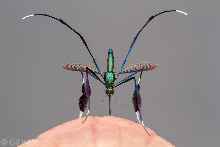
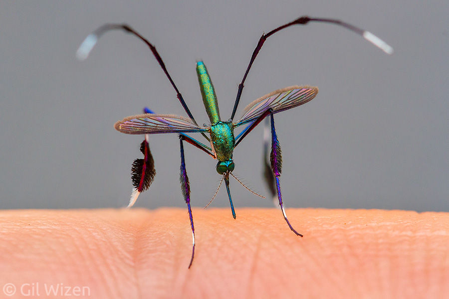
Beautiful bloodsucker. Wildlife Photographer of the Year, Invertebrate Behaviour category highly commended. A female mosquito (Sabethes sp.) in mid-bite. Amazon Basin, Ecuador

Beautiful bloodsucker. Wildlife Photographer of the Year, Invertebrate Behaviour category highly commended. A female mosquito (Sabethes sp.) in mid-bite. Amazon Basin, Ecuador

You can watch the part when it appears in the awards ceremony here (timestamped)

 
“Beautiful bloodsucker” is my personal favorite among my winning images, maybe because it took the greatest effort and longest time to produce (more on that later). This photo was released in September 2021 as a teaser for the Wildlife Photographer of the Year awards ceremony along with several other finalist images. Interestingly, upon release most media outlets chose to ignore the photo and omit it from their reports, however it eventually received exposure thanks to a BBC article that covered it extensively. The photo went viral online shortly after, as “the world’s most beautiful mosquito.”

“Now, come on. Come on! That’s the finest mosquito you’ve ever seen. It looks like a fantastic piece or art deco jewellery”

“Now, come on. Come on! That’s the finest mosquito you’ve ever seen. It looks like a fantastic piece or art deco jewellery”

You can also read the CBC article about “Beautiful bloodsucker” here or listen to my radio interview.

Why is this mosquito so flamboyant?
It is not clear why Sabethes mosquitoes have such beautiful metallic colors, but they are not the only ones. Other mosquitoes (for example Psorophora cyanescens, Toxorhynchites, etc’) have blue metallic scales covering their body. I discussed the fuzzy leg ornaments in a previous blog post; it is believed that they play a role in courtship, but because they are present in both sexes it is not well understood how they are being used.

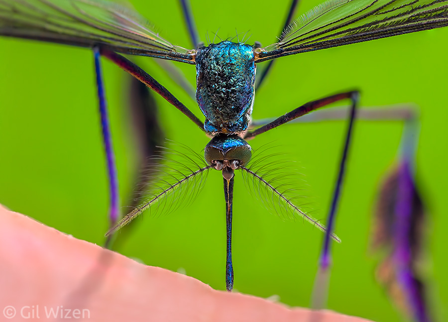
A closeup of a Sabethes mosquito in mid-bite, showing the beautiful scales covering its body

A closeup of a Sabethes mosquito in mid-bite, showing the beautiful scales covering its body

What are those curvy things above the mosquito’s head?
Those are the mosquito’s hind legs! All mosquitoes curve their hind legs upwards at rest, occasionally swinging them from side to side. These legs are covered with fine hairs and function as sensory organs to detect approaching threats. When an intruder (or a swatting hand) gets close, the legs detect the movement by changes in the air currents above the mosquito, prompting it to escape immediately by taking off.

What is the size of the mosquito?
Despite their exotic appearance, Sebethes mosquitoes are not too different in size from your typical mosquito. They are 4mm long, very tiny. The one photographed in my winning photo “Beautiful bloodsucker” is standing on my finger knuckle. Here is one photographed on my pinky finger for a better sense of scale.

Sabethes mosquito in mid-bite with pinky finger for scale

Sabethes mosquito in mid-bite with pinky finger for scale

Do you normally let mosquitoes bite you?
Not by choice, but when you are visiting a tropical rainforest it is bound to happen. Especially after rain, small water reservoirs in trees, epiphyte plants, or fallen leaves fill up and trigger female mosquitoes to go out looking for a blood meal before laying their eggs. I normally try to avoid getting bitten by wearing long sleeve clothes and putting some bug spray, but it is pretty impossible to avoid them entirely in the tropics. You can protect yourself as much as possible, but the moment you are distracted they will seize the opportunity to sneak up on you. Sabethes is the only mosquito towards which I am more forgiving. I love these mosquitoes, and every time I encounter them on my trips to South America I cheer with joy and hope that they come closer for a bite so I can take more photos of them. Am I a masochist? …maybe.

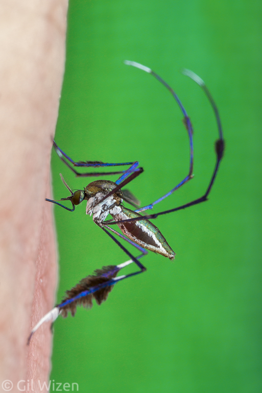
Sabethes mosquito in mid-bite. Note the swelling abdomen, filling with blood

Sabethes mosquito in mid-bite. Note the swelling abdomen, filling with blood

Isn’t this risky? Can this mosquito transmit any diseases?
It is important to remember that this is a wild animal, not a mosquito that was reared in a lab free of pathogens. There is definitely a component of risk here, as with all wildlife. These mosquitoes are important vectors of several tropical diseases, such as yellow fever and dengue fever, and perhaps other diseases as well. While taking the photo, I was bitten by this mosquito and several others, increasing the risk of contracting a vector-borne tropical disease. But I am still alive!

Is a bite from this mosquito painful?
Very. Every bite from a given species of mosquito feels a little different, mostly due to size and other morphological differences, but also thanks to differences in composition of the mosquito’s saliva. Sabethes mosquitoes are really something special. Not only do you feel them drilling into your skin, but it also leaves a painful sensation lasting hours and even days after the bite.

This Sabethes mosquito was photographed at the same location as the others, but it appears to be a different species with different leg ornaments.

This Sabethes mosquito was photographed at the same location as the others, but it appears to be a different species with different leg ornaments.

How long did it take you to get this photo?
About 4-5 years. I planned this particular photo composition for a long time. I have been encountering Sabethes mosquitoes for almost a decade and I knew I wanted something very specific. Little did I know that it would take an unbelievable amount of preparation and patience. These mosquitoes are extremely skittish and difficult to photograph well, especially in the constant heat and humidity of the rainforest. The mosquito responds to the tiniest of movements and to changes in light intensity. This means you must stay very still while attempting to photograph it, and also be prepared for the mosquito’s escape if using a flash. Fortunately, you are never alone with a single mosquito, because usually there are dozens of them hovering over your head. After carefully studying and observing the insect’s behavior for several years I was able to get a head-on, intimate photo of a female mosquito preparing to bite one of my finger knuckles. Even on the successful shoot itself it did not go smoothly the first time, and I had to keep trying for a couple of hours, all while getting bitten, until I finally got the photo I had in mind.

Why does this photo look like it was taken in a studio?
That is a great question that I received more than once. Indeed the background in “Beautiful bloodsucker” looks very plain and uniform to be considered in situ (in other words, a photo that was taken on site, in the subject’s natural habitat). However, I assure you that it was taken in the rainforest while I was visiting Ecuador. The background is simply the sleeve of my long hiking pants. After experimenting with different backgrounds on previous photography attempts, I chose this neutral background to emphasize the full spectrum of colors on the mosquito’s body.

I have seen many Sabethes mosquitoes over the years, but this female might be the prettiest so far

I have seen many Sabethes mosquitoes over the years, but this female might be the prettiest so far

Do you have any behind-the-scenes photos?
I do not, but maybe this it for the best. Almost every time I attempted to photograph these mosquitoes I was surrounded by a swarm of them. It was very annoying and I was frustrated from getting bitten. I bet it would look horrible on camera.

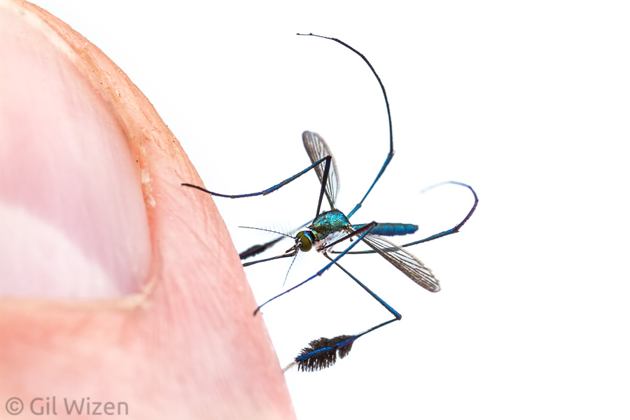
Sabethes mosquito biting my thumb

Sabethes mosquito biting my thumb

Do you think this photo will change people’s general view on mosquitoes?
I am a pretty realistic guy, so I do not expect my photo to make people fall in love with mosquitoes. My hope is that it will make people pause and look before they automatically swat a mosquito, and maybe appreciate and beauty and structural complexity of these animals.

We like the photo! Can we buy a print from you?
Of course. You can get a wall print of “Beautiful bloodsucker” directly from the Natural History Museum’s shop at affordable rates. If you need something different, contact me and I will do my best to assist you.

If you have any questions about my photo that do not appear in this post, feel free to leave them in the comments. I will do my best to answer them.

To read part 1 about “The spider room”, click here.
To read part 2 about “Bug filling station”, click here.
To read part 4 about “Spinning the cradle”, click here.
To read part 5 about what to expect when entering a photo competition, click here.

Wildlife Photographer of the Year Q&A: Bug filling station, Behaviour: Invertebrates highly commended

We are continuing our series of Q&A posts about my Wildlife Photographer of the Year winning images, and this time I will be reviewing Behaviour: Invertebrates highly commended: Bug filling station.

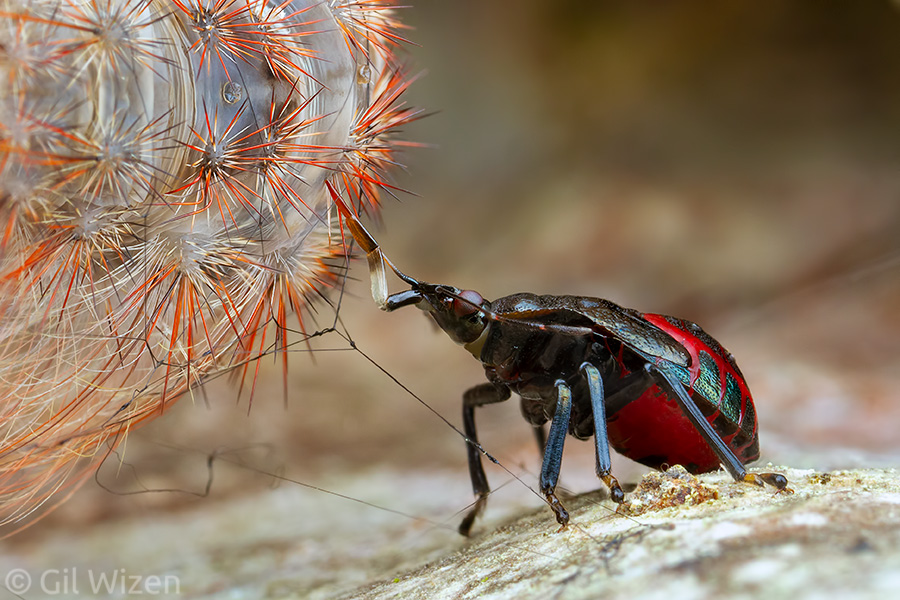
Bug filling station. Wildlife Photographer of the Year 2021, Invertebrate Behaviour category highly commended. Predatory stink bug nymph (Euthyrhynchus floridanus) feeding on a moth caterpillar. Mindo, Ecuador

Bug filling station. Wildlife Photographer of the Year 2021, Invertebrate Behaviour category highly commended. Predatory stink bug nymph (Euthyrhynchus floridanus) feeding on a moth caterpillar. Mindo, Ecuador

You can watch the part when it appears in the awards ceremony here (timestamped)

 
Out of my winning images, “Bug filling station” received the least attention and was skipped by many of the reporting media outlets. I think it is a shame, because it tells an interesting story of opportunistic survival.

“It’s a gruesome scene but it’s a remarkable piece of behavior”

“It’s a gruesome scene but it’s a remarkable piece of behavior”

What is so special about this photo?
The photo shows a small bug nymph feeding on a much larger moth caterpillar that was in preparations for pupating on a tree trunk. However, there is more depth to this story. The caterpillar is most likely a species of a tiger moth, which are characterized by having thick barbed hairs or spikes for protection against predators and parasitoids. As you can see, it didn’t really help the caterpillar in this case, for two reasons. First, the caterpillar is resting inside a very thin and poorly constructed cocoon, these are the black silk threads that can be seen in the photo. This cocoon is spacious and open because the cloud forest is a very wet environment. A typical cocoon with high-density spun silk will absorb rainwater and drown the pupa inside, whereas an open cocoon drains water better. Even though it is a thin cocoon, the caterpillar is still trapped inside and cannot leave. Second, pupating caterpillars are helpless and cannot defend themselves, as they lose the ability to walk prior to pupation, along with most of their senses. Therefore, the caterpillar is essentially defenseless at this stage, and indeed many predators and parasitoid insects seize this opportunity to attack.
Now the small bug nymph enters the picture, and decides to take advantage of the immobilized caterpillar by piercing its body with its proboscis and sucking its juices while the caterpillar is still alive. It is a great opportunity for the bug, because it can stay next to the caterpillar and feed as long as it wants or needs, without worrying about the prey escaping or the food supply running out.

Can you elaborate more about the bug’s mouthparts? I can’t understand what I am looking at.
The hemipteran proboscis is actually a complex system of mouthparts for sucking. The liquid food travels inside the narrow tube seen at the top of the mouthparts complex. This tube consists of the “jaws”; the elongated mandibles and maxillae are layered and arranged as a feeding tube. The folded part seen at the bottom is called labium (lower lip), and functions as a sheath to keep the mouthparts packed tightly together. During feeding this sheath is pushed backwards to expose the tip of the feeding tube and allows the bug to “bite” and start drinking.

What is the size of the bug?
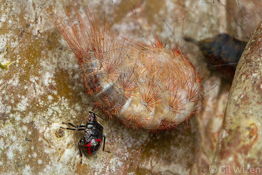
Body length was 8mm. Judging by the size of the wing buds I would say it is two stages (=instars) away from becoming an adult.

A slightly different view of the bug filling station. The Euthyrhynchus floridanus nymph is very small compared to the huge moth caterpillar!

A slightly different view of the bug filling station. The Euthyrhynchus floridanus nymph is very small compared to the huge moth caterpillar!

Do you have any behind-the-scenes photos?
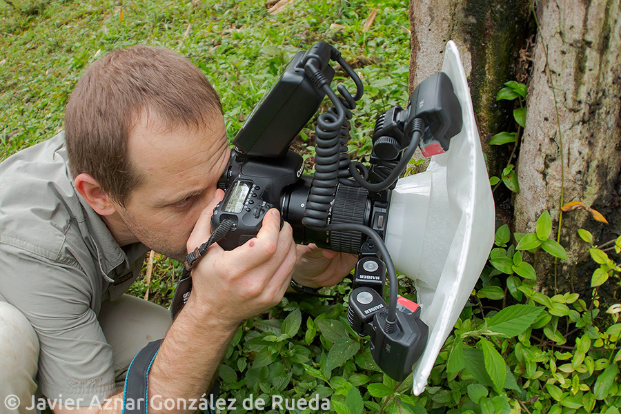
I usually travel alone, but surprisingly in this case I do! I spent a few days in the cloud forests of Mindo, Ecuador together with my friend Javier Aznar, taking photos of the beautiful arthropod fauna there. He was kind enough to take my photo.

Me photographing in Mindo, Ecuador (photo courtesy of Javier Aznar)

Me photographing in Mindo, Ecuador (photo courtesy of Javier Aznar)

What else can you tell us about this bug?
The species is the Florida predatory stink bug (Euthyrhynchus floridanus). It is a monotypic species, in other words it is the only species in its genus. It has a wide distribution in southeastern United States and northern Latin America. In contrast to most members of its family Pentatomidae, this species is carnivorous and considered beneficial. It seems to enjoy feeding on many plant pests, as well as other small insects. Interestingly, this species is also gregarious, sometimes attacking prey in groups, although in my case no other nymphs were present in the area. The adult bugs display high color polymorphism, with variable red or orange patches on a metallic dark blue body.

We like the photo! Can we buy a print from you?
Of course. Contact me and I will do my best to assist you.

If you have any questions about my photo that do not appear in this post, feel free to leave them in the comments. I will do my best to answer them.

To read part 1 about “The spider room”, click here.
To read part 3 about “Beautiful bloodsucker”, click here.
To read part 4 about “Spinning the cradle”, click here.
To read part 5 about what to expect when entering a photo competition, click here.

Wildlife Photographer of the Year Q&A: The spider room, Urban Wildlife category winner

Recently I was honored to have four of my photos commended in the prestigious Wildlife Photographer of the Year photo competition organized by the Natural History Museum in London. This is a major achievement for me, and not something that I take lightly. A lot of hard work, dedication, and patience got me to this point. Most photographers spend years trying to get a single photo recognized in the competition, usually without success. To have four entries selected as finalists, with two category winners, is not something I expected even in my wildest dreams. The attention from the press and the general public after the awards ceremony and the winners announcement (watch it here) was overwhelming and sometimes exhausting (especially in the case of the photo “The spider room”). Now that things have calmed down a little, I decided to dedicate a few posts to the competition; to answer some of the repeating questions from people, and provide a bit of the background story for each of my winning photos.

We start off this series of posts with Urban Wildlife category winner: “The spider room”. First of all if you have not read the full story behind this photo, feel free to head over to this post.

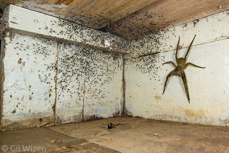
The spider room. Wildlife Photographer of the Year 2021, Urban Wildlife category winner. Phoneutria fera and its babies under my bed. Amazon basin, Ecuador

The spider room. Wildlife Photographer of the Year 2021, Urban Wildlife category winner. Phoneutria fera and its babies under my bed. Amazon basin, Ecuador

You can watch the part when it appears in the awards ceremony here (timestamped).

Out of my winning images, this is probably the photo that made the most impact. The public response to it was phenomenal. It went viral immediately after the awards ceremony, attracting comments from thousands of people. I got literally hundreds of messages and questions about it over social media. It seems that people either like this photo, or really, REALLY hate it. The interesting thing is that either way, people have an opinion about it. They talk about it. It’s a conversation starter. I couldn’t ask for a better result. There is also a story behind the submission of this photo to the competition that I will mention later.

“Seriously... This was under Gil Wizen’s bed, I’m sure it might send a shiver down your spine, but when I tell you that it was a Brazilian wandering spider, a very large animal and one of the most venomous spiders in the world, you’d be more worried”

“Seriously… This was under Gil Wizen’s bed, I’m sure it might send a shiver down your spine, but when I tell you that it was a Brazilian wandering spider, a very large animal and one of the most venomous spiders in the world, you’d be more worried”

Ok, spill out the truth. Is it real?
Yes.

What is the size of the spider? It looks huge!
This is an adult female Phoneutria fera, or Brazilian wandering spider. It is one of the largest araneomorph (non-tarantula) spiders in the world. The spider can easily cover an adult human’s hand with its leg span, which is almost 6 inches or 15 centimeters. The lens used to capture the photo makes it look bigger (forced perspective).

What is this “forced perspective”?
Forced perspective is an optical illusion that makes an object appear physically different (larger, smaller, closer, or farther) than it actually is. In this case I used a short focal length wide-angle lens, photographing the spider under my bed from a very short distance to make it appear larger in the frame while still retaining most of the details of the background.

Don’t spiders have 8 legs? I only see 6!
Spiders indeed have eight legs. This spider is not missing any legs, but it holds the two front pairs closely together, making it look like it is a single pair of legs.

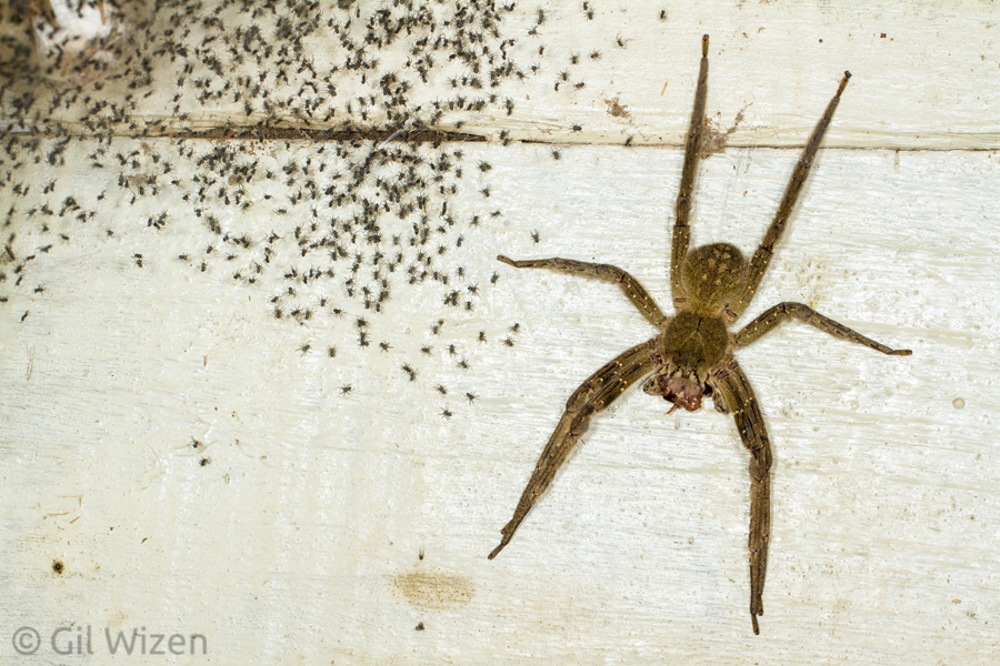
A wandering spider (Phoneutria fera) female guarding her babies. Amazon Basin, Ecuador

A wandering spider (Phoneutria fera) female guarding her babies. Amazon Basin, Ecuador

What is that thing on the floor next to the spider?
The spider is feeding on a cockroach, so right under it there is a cockroach leg that it discarded. However, since the online version of the photo is usually of low resolution, most people mean the black area on the floor in front of the spider – that is simply a hole in the floorboard.

How venomous is this spider?
To quote from wandering-spiders.net: “Phoneutria venom contains a wide variety of peptides and proteins including neurotoxins, which act on the ion channels and chemical receptors of the neuromuscular systems of insects and mammals.” This means the venom has the potential to cause excitatory symptoms such as salivation, muscle spasms, loss of consciousness, loss of control over muscles, priapism (yup, you read that right), and in some cases even death.

Oh no! Banana spider aka Brazilian wandering spider (Phoneutria fera) in my kitchen!

Oh no! Banana spider aka Brazilian wandering spider (Phoneutria fera) in my kitchen!

Where was this photo taken? I am worried!
The spider room photo was taken in a biological research station in Ecuador. Phoneutria spiders are only found in the tropical regions of Latin America.

How did you not set the whole room on fire?? What’s wrong with you, look at this thing!
And why would I do that? The spider doesn’t know it is in someone’s room. It doesn’t even know what a human is. Allow me to quote myself: We DO NOT burn houses just because a spider happened to walk in. It’s absurd. Just because a spider found its way into your house, doesn’t mean it’s going to go after you. Spiders are constantly busy surviving, they have no time for us. If you find a spider at home, please kindly escort it out. The spider will thank you, and both of you will be happy. No need to cause property damage and possibly hurt yourself and others in the process.

It doesn’t look like any bed that I’ve ever seen. Can we see the bed?
Yes you can! Just don’t expect too much.

The bed where Phoneutria fera was found (photo courtesy of Alex Shlagman)

The bed where Phoneutria fera was found (photo courtesy of Alex Shlagman)

This photo isn’t mine. It was taken by my colleague during our previous stay at the site in 2007 and the room has changed considerably since then, but it’s the same bed.

Do you have any behind-the-scenes photos?
Unfortunately I was alone during the encounter with the spider, so I have no behind-the-scenes photos. However, I can try to communicate the experience. After figuring out that the source for the baby spiders in my room was under the bed, I decided to crawl under it to take a closer look. Someone on twitter posted this image, either directly or indirectly connected to my spider photo, and it encapsulates the scene very well:


When I looked under the bed, this is what I saw:

Phoneutria fera under my bed. Amazon basin, Ecuador

Phoneutria fera under my bed. Amazon basin, Ecuador

And one look was all I needed. I immediately knew which kind of photo I wanted to take.

Weren’t you scared to get so close to the spider?
I was not scared to photograph the spider from up close, because it was busy feeding and did not pay attention to me. However, after photographing I decided to relocate it outside, and this involved moving the spider. I was a little concerned because this spider is extremely fast and defensive, so I moved slowly and used extreme caution.

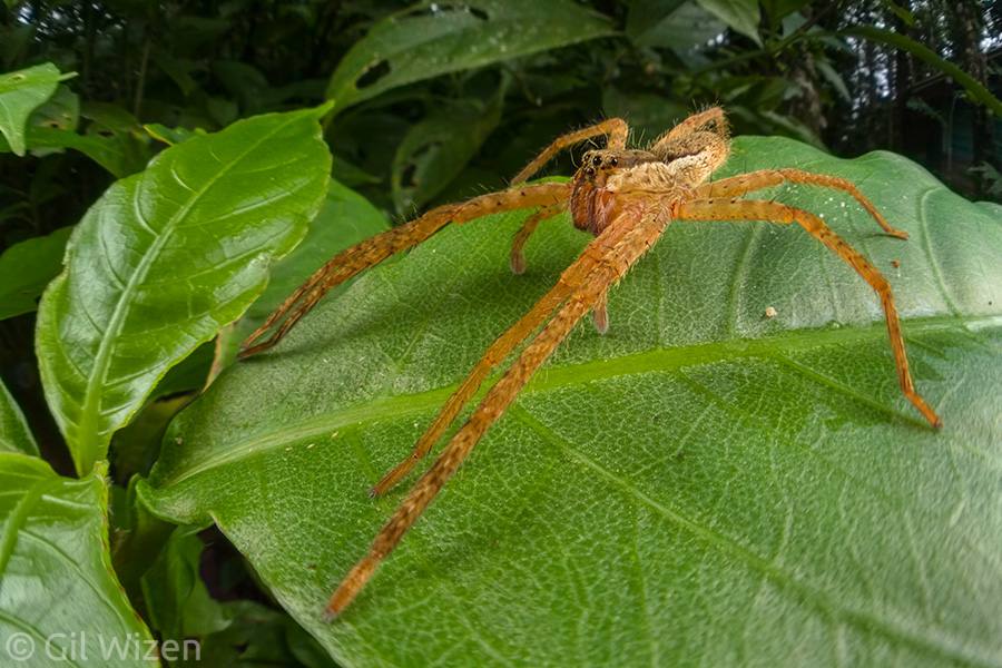
Closeup of a wandering spider (Phoneutria boliviensis) resting on a leaf in the Ecuadorian Amazon. I got very close when taking this photo and the spider could not care any less.

Closeup of a wandering spider (Phoneutria boliviensis) resting on a leaf in the Ecuadorian Amazon. I got very close when taking this photo and the spider could not care any less.

How did you move the spider outside?
There is a simple trick for catching spiders by placing a cup over the arachnid and sliding a piece of paper underneath. It works with any spider, large or small, tarantula or araneomorph spider. This is what I did with the Brazilian wandering spider before carrying it outside to release.

You said you relocated the spider outside, but what about all the babies?
The babies were left untouched. After hatching from the egg sac, baby spiders need very little attention. In some spiders the mothers stay close to the babies to protect them, but the truth is they are independent and can take care of themselves. That is why they were already dispersing around in the room. In addition, the baby wandering spiders have tiny fangs and physically cannot bite humans, so they are harmless.

Don’t spiders go back to their nest if moved away from it? Did the spider return?
After it was released the spider did not return to the room. However, this question is justified because I have heard more than one account where a wandering spider was relocated and showed up in the same place the day after. It is possible that the spider can find its way back following chemical cues. Silk may contain important information about the individual spider that placed it, and this information can be used by the same animal or other spiders for tracking.

Did you have any similar encounters with these spiders?
I encounter members of genus Phoneutria almost every time I visit Latin America. I always get startled at first because it is a very large spider, but then I continue to observe them without worries. They are interesting animals with an important role in their habitat and we should treat them with respect.

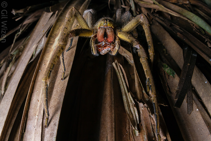
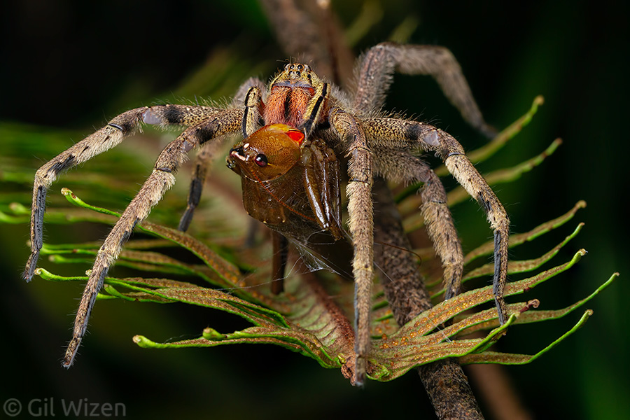
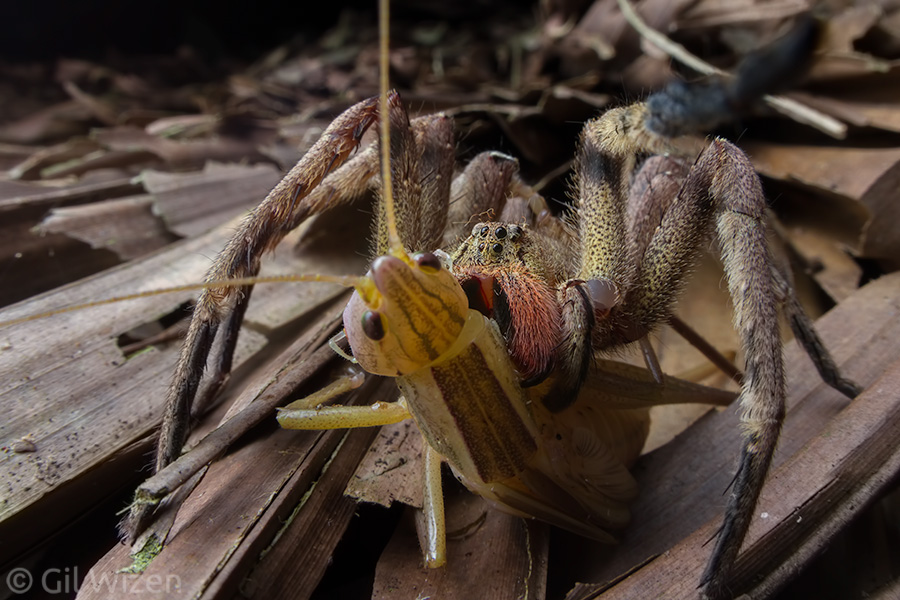
Wandering spider (Phoneutria depilata) preying on a katydid. Photographed on the thatched roof of a cabin in Colombia.

Wandering spider (Phoneutria depilata) preying on a katydid. Photographed on the thatched roof of a cabin in Colombia.

Wandering spider (Phoneutria depilata) preying on a katydid. Limón Province, Costa Rica

Wandering spider (Phoneutria depilata) preying on a katydid. Limón Province, Costa Rica

We like the photo! Can we buy a print from you?
Of course. Contact me and I will do my best to assist you.

Can I use your photo for my super funny meme? Please!
This has already happened even before the photo won in the competition.
I had a feeling that the photo would go viral after the winners announcement, and expected the internet to have a field day using it for memes. As long as the memes are civil, do not call for violence, damage of property, or the unnecessary killing of spiders – I am fine with it.

What is the story behind the submission that you mentioned in the beginning of the post?
As mentioned in my award reception speech for “The spider room”, I actually had no intention to submit this photo to the competition. My plan was to submit another photo of a wandering spider preying on a katydid, however my good friend Ellen Woods, who encouraged me to enter my work to the competition in the first place (something I will discuss in a later post in this series), insisted that I submit this particular photo in the Urban Wildlife category. And she was spot on! So the way I see it, this is more her win than mine.

Wandering spider (Phoneutria depilata) preying on a katydid. Photographed in Colombia. This was the photo I initially planned to submit to the competition. Looking back, it would probably not have been picked up as a finalist.

Wandering spider (Phoneutria depilata) preying on a katydid. Photographed in Colombia. This was the photo I initially planned to submit to the competition. Looking back, it would probably not have been picked up as a finalist.

Why did you submit this photo in the “Urban Wildlife” category, when it shows a research station close to the rainforest and has nothing to do with being urban?
I admit I hesitated to submit the photo because of this. However upon careful inspection of the category’s definition in the competition, the text reads: “Across the world, humans have created new habitats. Many animals have adapted to these built environments, some more successfully than others. These images focus on the magic of the commonplace, the surprise of the unexpected or the wonder of the normally unseen.” This means that the category is intended for photos of animals adapting to any human-made environment. Not necessarily just cities, even if it is the first thing that comes to mind when hearing the word “urban”.

If you have any questions about my photo that do not appear in this post, feel free to leave them in the comments. I will do my best to answer them.

To read part 2 about “Bug filling station”, click here.
To read part 3 about “Beautiful bloodsucker”, click here.
To read part 4 about “Spinning the cradle”, click here.
To read part 5 about what to expect when entering a photo competition, click here.
Bachia lizards – look, no hands!

Legend tells the story of Oedipus, who faced the sphinx guarding the gates to the city of Thebes. To enter, the monster presented him with a riddle:
“Which creature has one voice and yet becomes four-footed and two-footed and three-footed?”
Oedipus was able to solve the sphinx riddle, granting him entrance to the city. He later became the king of Thebes and married his own mother – but that’s another story.
The answer to the sphinx riddle can be quite intuitive if you stop to think about it, but what if I told you there are animals that fit that description quite easily? It is with my own eyes that I have seen arachnids staring their life with eight legs, losing some of them during growth, and then growing them back like nothing happened in the amazing process of regeneration. Some hemimetabolous insects and even amphibians can do it just as well. And what if I told you there is an animal that is born with legs, and by the end of its life it loses all of them but one? Introducing Bachia, a genus of strange long-bodied lizards.

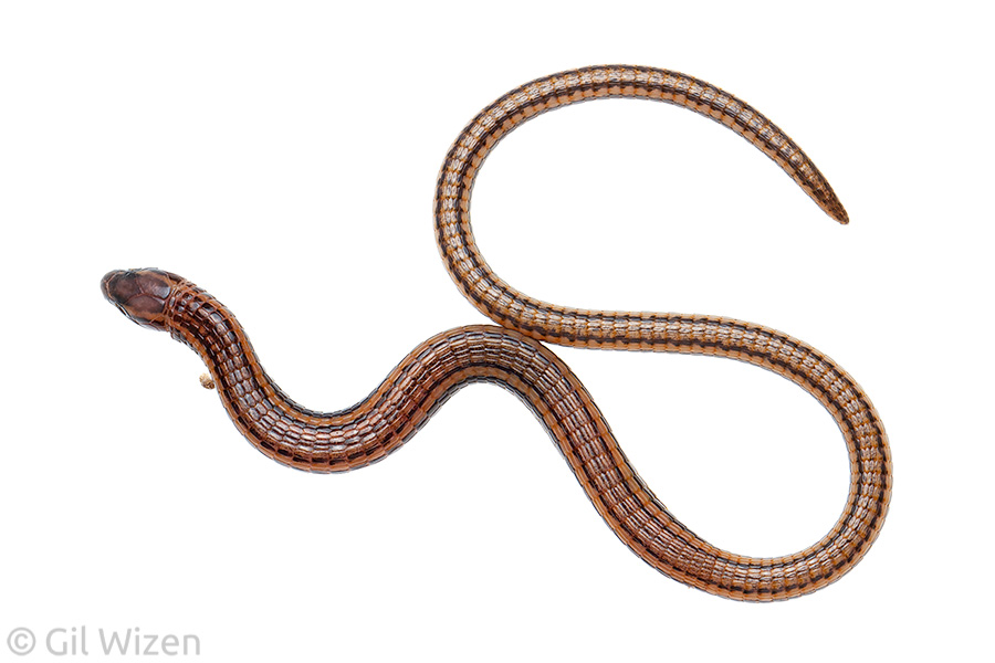
Stacy's bachia (Bachia trisanale), a limbless microteiid lizard from the Ecuadorian Amazon

Stacy’s bachia (Bachia trisanale), a limbless microteiid lizard from the Ecuadorian Amazon

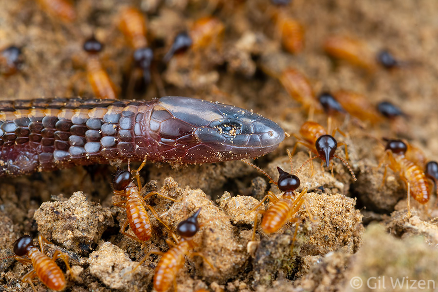
The adult Bachia lizards resemble snakes, having an elongated body and reduced limbs. They have small eyes and no external ear openings. These are adaptations for a subterranean lifestyle, as these lizards spend most of their time moving through the leaf litter and in the soil looking for their favorite food – soft-bodied insects. They usually hunt termites and ant brood in underground nests. Occasionally they make their way into decomposing wood, where they will not hesitate to snag a juicy beetle larva if the opportunity presents itself. I encountered my first Bachia while it was hunting termites moving on a trail.

Stacy's bachia (Bachia trisanale) hunting termites

Stacy’s bachia (Bachia trisanale) hunting termites

Stacy's bachia (Bachia trisanale) hunting termites

Stacy’s bachia (Bachia trisanale) hunting termites

Termite soldiers surrounding the intruding Bachia lizard to defend their colony workers on the trail

Termite soldiers surrounding the intruding Bachia lizard to defend their colony workers on the trail

Closeup on a Bachia's head, showing small eyes and no external ear opening - adaptations for a subterranean lifstyle.

Closeup on a Bachia’s head, showing small eyes and no external ear opening – adaptations for a subterranean lifstyle.

Bachia are not to be confused with other snake-like lizards such as limbless skinks, slowworms, glass lizards, or amphisbaenids, all members of other groups. Bachia lizards belong to family Gymnophthalmidae, also known as microteiids or spectacled lizards. Their closest relatives are the skittish whiptail lizards. The name “spectacled” refers to their transparent eyelids, allowing these lizards to see even when their eyes are closed. Most members of the family are normal looking lizards, like this common root lizard (Loxopholis parietalis).

Juvenile common root lizard (Loxopholis parietalis). Amazon Basin, Ecuador

Juvenile common root lizard (Loxopholis parietalis) from the Ecuadorian Amazon

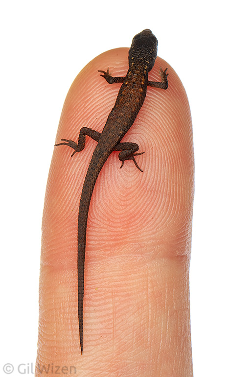
As the name suggests, microteiid lizards are generally small. How small? Very small.

Juvenile common root lizard (Loxopholis parietalis) on finger for scale

Juvenile common root lizard (Loxopholis parietalis) on finger for scale

Ok, I’m cheating here a little, after all this is a juvenile specimen. But generally speaking, a microteiid lizard can sit comfortably in the palm of your hand, and you will still have room for two or three more lizards.

Portrait of Stacy's bachia (Bachia trisanale)

Portrait of Stacy’s bachia (Bachia trisanale)

The genus Bachia contains about 30 species, many of which are endemic. The species I have encountered the most is Bachia trisanale, it is one of the more common species with a wide distribution in South America, occurring in Ecuador, Peru, Bolivia, Colombia, and Brazil.

Stacy's bachia (Bachia trisanale), full body view

Stacy’s bachia (Bachia trisanale), full body view

One of the most interesting aspects of Bachia lizards is their limbs. All species have a long body with very small limbs. Members of the genus can be easily divided into groups by their limb structure and the level of reduction in the hindlimbs. In many species the hindlimbs are extremely reduced to tiny hooks or absent altogether. Few species, like the Bachia trisanale appearing in this post, lack hindlimbs and show digit reduction in the forelimbs as well. Interestingly, limbs can also be lost throughout the lizard’s lifetime. Although a leg can be lost following an injury, it can also happen due to tissue erosion caused by the lifestyle of digging and burrowing through coarse soil containing clay particles. The hind legs (if present) usually go first, shrinking to tiny knobs or disappearing completely. The stronger forelimbs are eventually eroded as well, first the tiny digits, and then the remainder of the leg. Judging by several specimens that I have encountered, the forelimb loss is even a directional change – usually it is the right forelimb that disappears first, followed by the one on the left side. It is common to find old Bachia lizards with only one limb! Usually by that time the remaining leg has lost its digits entirely, and looks like a small stub.

Stacy's bachia (Bachia trisanale), closeup on its head and stubby foreleg

Stacy’s bachia (Bachia trisanale), closeup on its head and stubby foreleg

Surprisingly this has no negative effect on the lizard’s locomotion. One might even argue that losing the limbs makes it more terradynamic, allowing it to “swim” freely in the substrate with no drag.

Bachia trisanale with only one foreleg

Bachia trisanale with only one foreleg

So when you are feeling down, remember that there are small lizards that look like a noodle, diving face first into the dirt after pesky termites and ants. And they do it with no hands!

Ending 2018 with a bang

Let’s go straight to the punchline: 2018 was a successful year for me. This is important because I am posting this late into 2019, so I had the time to reflect on the passing year’s events.
I was debating if I should write a summary post for 2018 at all. These things get old quickly, especially around the last two weeks of December, when such posts pop up everywhere. Even though I know I am expected to post my favorite photographs from last year, the truth is that I have not been particularly busy taking photos. Instead I have been focusing on other projects, giving invited talks and doing public outreach, as well as adding content to this website. In addition, there were a few notable events this year that helped shaping me as a professional and as a person. So if you want to know what I have been up to in 2018, read on.

Teaching about whip spiders at the Royal Ontario Museum

Teaching about whip spiders at the Royal Ontario Museum

Opinion piece about giving away free photos,
and the rise to fame (not really)

One of the first posts I wrote in 2018 was aimed at people asking for free work. It joined other similar posts (examples are here and here), and to be honest I did not expect to receive any response to it. I merely posted the article so I can go back and refer people to it whenever I am faced with such requests. As many creative freelancers know, being asked to work or provide service for free is far too common. Apparently that post hit close to home because it received echo from fellow photographers and artists, and was later picked up by PetaPixel, one of the biggest photography-related news websites.
The piece featured on PetaPixel spread like wildfire and triggered some interesting discussions. On one hand, not everyone liked what I wrote, and some users even attacked me for it. On the other hand, I found myself discussing important topics like rates and copyrights with other, more experienced professionals. Even though this rant has been years in planning, I admit the post was written in the heat of the moment, and as such it is not a well-worded text. Now that I think about it, it is clear that I could have done a better job. That being said, I have no regrets whatsoever; the post is justified. If anything, having it available on my website serves as a deterrent for anyone who behaves inappropriately and disrespectfully towards creators by asking for free work.

Trip to Colombia

In February I went on a short assignment in Colombia, to photograph a tree. I know, right? It sounds simple and too good to be true, but in reality finding trees that fit what the client had in mind was challenging, not to mention it was not always possible to shoot the tree in the requested high resolution for the project. This trip also gave me a chance to test the Laowa 25mm f/2.8 2.5-5X Ultra Macro lens, and I am happy I did, because it proved to be an outstanding competitor to the renowned Canon MP-E 65mm f/2.8 1-5x macro lens. Laowa is quickly establishing themselves as one of the most interesting lens companies out there, and I am looking forward to see what other innovations they have up their sleeve!

Searching for trees. Anyone seeing any trees around?

Searching for trees. Anyone seeing any trees around?

Tree covered in climbing epiphytes. Is this the perfect tree for the job? Close to perfect, but unfortunately, it was not good enough.

Tree covered in climbing epiphytes. Is this the perfect tree for the job? Close to perfect, but unfortunately, it was not good enough.

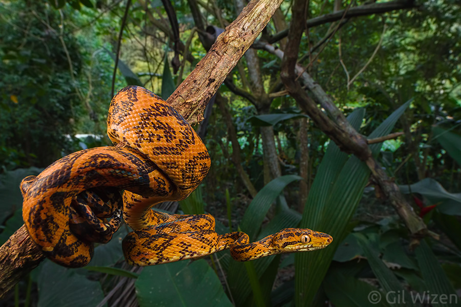
Central American tree boa (Corallus ruschenbergerii). One of the benefits of walking around checking out trees, is finding tree-inhabiting animals.

Central American tree boa (Corallus ruschenbergerii). One of the benefits of walking around checking out trees, is finding tree-inhabiting animals.

Katydid nymph showing intense pink coloration or erythrism - a rare phenomenon that is occasionally seen in katydids, in which a genetic mutation causes an absence of the normal green pigment and the excessive production of pink pigment.

Katydid nymph showing intense pink coloration or erythrism – a rare phenomenon that is occasionally seen in katydids, in which a genetic mutation causes an absence of the normal green pigment and the excessive production of pink pigment.

Fairy wasp (family Mymaridae) ovipositing in membracid treehopper egg mass. One of the best finds during this trip (watch this space for an upcoming post about it). This tiny wasp is only 0.6mm in length! Photographed using the Laowa 25mm lens.

Fairy wasp (family Mymaridae) ovipositing in membracid treehopper egg mass. One of the best finds during this trip (watch this space for an upcoming post about it). This tiny wasp is only 0.6mm in length! Photographed using the Laowa 25mm lens.

Trip to Ecuador

In May I returned to my beloved site in Ecuador, this time it wasn’t really to get work done but was more of an escape to spend my birthday (ok, who am I kidding, I still worked during that visit…). I often feel miserable around my birthday and during holidays because many times I have no one to spend them with, and it seemed like a good idea to pass this time in a place that I love. More than anything I needed to clear my head before taking the role of a spider wrangler (up next). This trip ended up being very peaceful and relaxing, somewhat lonely, but still satisfying. I was able to finally find one of my target species for the area, the seasonal jewel scarab, Chrysophora chrysochlora.

Ecuadorian jewel scarab (Chrysophora chrysochlora)

Ecuadorian jewel scarab (Chrysophora chrysochlora)

Bird eater tarantula (Theraphosa sp.). I was extremely happy to encounter this giant for the first time in the wild, and add it to the species checklist for my site.

Bird eater tarantula (Theraphosa sp.). I was extremely happy to encounter this giant for the first time in the wild, and add it to the species checklist for my site.

Male orchid bee (Euglossa intersecta) in mid-flight. This photo is not perfect in many ways: it is dark, the composition is not ideal, and it is also slightly cropped. But I achieved something very difficult here - taking a wide angle macro and freezing an orchid bee in flight. Those bees are fast!

Male orchid bee (Euglossa intersecta) in mid-flight. This photo is not perfect in many ways: it is dark, the composition is not ideal, and it is also slightly cropped. But I achieved something very difficult here – taking a wide angle macro and freezing an orchid bee in flight. Those bees are fast!

Male orchid bees (Euglossa sp.) collecting fragrant compounds from a moss patch

Male orchid bees (Euglossa sp.) collecting fragrant compounds from a moss patch

Baby wandering spider (family Ctenidae)

Baby wandering spider (family Ctenidae)

Spiders exhibition at the Royal Ontario Museum

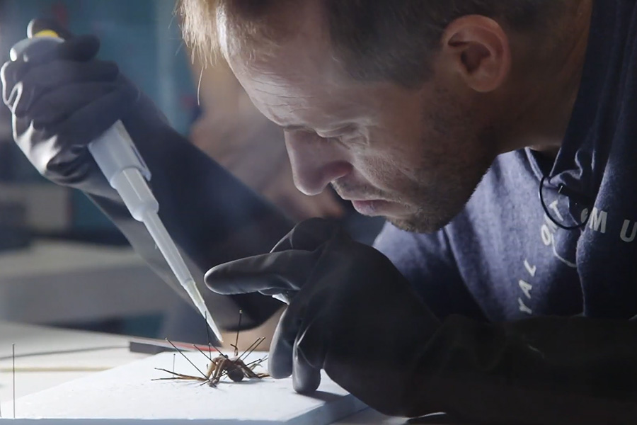
And then, immediately after returning from Ecuador, I was recruited to the Royal Ontario Museum to work on their “Spiders: Fear & Fascination” exhibit. This is something I knew I wanted to do ever since I heard about this exhibit in December 2016. It was a long wait but I was finally in! I worked full-time as the spider wrangler for seven months at the museum, along with the awesome Mateus Peppineli. The role involved mainly taking care of spiders displayed in the gallery and in the live room, but also delivering presentations to the museum visitors. These presentations were open for whatever came in mind, so I could include information about different arachnids, not just spiders (as can be seen in the photo opening this post). A key component of the presentations was performing live venom milking from spiders and scorpions in front of an audience (you can watch a venom milking presentation in this video). This is something that requires some explanation – the idea was to extract venom from live animals that were sedated, and do it in a way that would not harm the animals and allow for future extraction of venom later on.

Milking venom from a fishing spider at the Royal Ontario Museum. Screenshot from TFO video

Milking venom from a fishing spider at the Royal Ontario Museum. Screenshot from TFO video

This is something I got trained for about a decade ago in Israel, I felt so lucky to be able to not only do this again, but also to present it to other people. In this regard I believe this ROM exhibit was one of a kind. Sure, the original exhibit coming from Australia also had a venom milking component, but I think the exhibit is really as good as the person running the show. I don’t know if there is any other place that has the unusual combination of someone passionate about arachnids, has over 20 years worth of expertise keeping and designing public displays for them, AND is also trained in venom extraction. And this is exactly why this exhibit made it well; people hoarded to see it, many coming back several times for more. Not only it gave me enormous satisfaction to see that people are interested in spiders, but also it showed me that without doubt there is a desire to see a similar permanent exhibit in Toronto, maybe on a larger scale. And I needed that affirmation.

Presenting spiders to an audience at the Royal Ontario Museum. Screenshot from Curiosity in Focus video

Presenting spiders to an audience at the Royal Ontario Museum. Screenshot from Curiosity in Focus video

I met and talked with thousands of people. Many of them came to me wishing to learn more about spiders they saw in their own house or backyard. Children found in me someone who is on their side, because I could understand and relate to their passion for insects and spiders. Some people confessed that I changed their point of view and helped them conquering their fear of spiders. Young visitors told me that watching me working has inspired them to pursue higher education and a career in science. And sure, it was nice being in the spotlight for a little while.

Some screenshots from TV appearances, representing the ROM's "Spiders: Fear & Fascination" exhibit

Some screenshots from TV appearances, representing the ROM’s “Spiders: Fear & Fascination” exhibit

I enjoyed being the ambassador for arachnids on TV and social media. People started recognizing me on the street. Suddenly, I had fans. I got hugs. So many hugs. Some visitors stayed in touch and became close friends. I want to thank everyone who helped making this exhibit into a reality, and all the visitors who gave it a chance and came to see it. Now that the dust has settled I can honestly say that working at the spiders exhibit was one of the best experiences of my life. How do I fill the void that is left, that is the question that bothers me the most right now.

I will never forget this "finger fencing" moment. Screenshot from CP24 Live at Breakfast

I will never forget this “finger fencing” moment. Screenshot from CP24 Live at Breakfast

Social media (Instagram)

As a part of my role at the museum, I decided to join the social media platform Instagram. I did it as an experiment, to see how well I can perform there. If you know me, then you know that I find Instagram useless and crippled compared to the other social media streams, because of the inability to post clickable links or share posts from other users. My initial thought was to share behind-the-scenes moments from my work at the museum, and indeed the very first posts were exactly that. However, I quickly learned that this does not always work well, I did not always have the time to post while working, and if I have to be completely honest, my phone camera is total garbage. Nevertheless, I persisted, posting the same stuff that I post on my other accounts, and people started following. So maybe it is not so bad… I am still a shy noob on Instagram and don’t interact much, but if you remember my post from 2016, I said the exact same thing about Twitter, and today it has become my preferred social media platform. So who knows. I’m ready for an adventure!

Collaborating with Daniel Kwan (Curiosity in Focus)

During my time as the Royal Ontario Museum’s spider wrangler I was fortunate to meet Daniel Kwan, a talented individual who was responsible for running educational programs at the museum as well as producing video content about the different museum exhibits and collections. Since the opening of the spiders exhibit we collaborated many times, producing videos showing live arthropods and their behaviour, as well as activities performed in the venom lab.


Daniel also has several side projects that he runs. Many of them are related to his big passion – games, but he also hosts a couple of podcasts. One of them, Curiosity in Focus, shines a spotlight on people with interesting professional lifestyles and backgrounds, and covers many different fields like science, history, archeology, design, and fiction. Ever since the day we met he has been trying to get me on the podcast, until we finally found the time to sit down one evening and record an episode. So if you like to know about more about me, feel free to check it out here. We chatted about my role as the spider wrangler at the ROM, my military service in Israel, and shared some stories from entomology-related fieldwork. This was so much fun. I am definitely looking forward to future collaborations with Daniel.
Regardless of this episode, I have nothing but good things to say about this podcast. This is Daniel’s own brainchild, he is very passionate about it, and it shows. He always finds amazing people and interesting topics to talk about. One of my personal favorite episodes is a chat with Kevin Rawlings, Canada’s northernmost resident. Check it out if you have a moment. You won’t regret it.

Gilwizen.com in 2018

Due to my trips and later employment at the museum, my posting routine on the blog was inconsistent. I posted only 14 stories on the blog this year, a huge drop compared to 30 posts in the previous year. I am happy with the posts I got to share, but I would like to write more. Even more surprising was to see old posts resurfacing on social media and attracting new readers, notably this one, this one, this one, and… this one? Hmm. Some of these are very old and there is no doubt my writing style has changed over the years. I am tempted to go back and rewrite some of them to make it easier and more interesting to read, but I know that something would be lost if I do so.

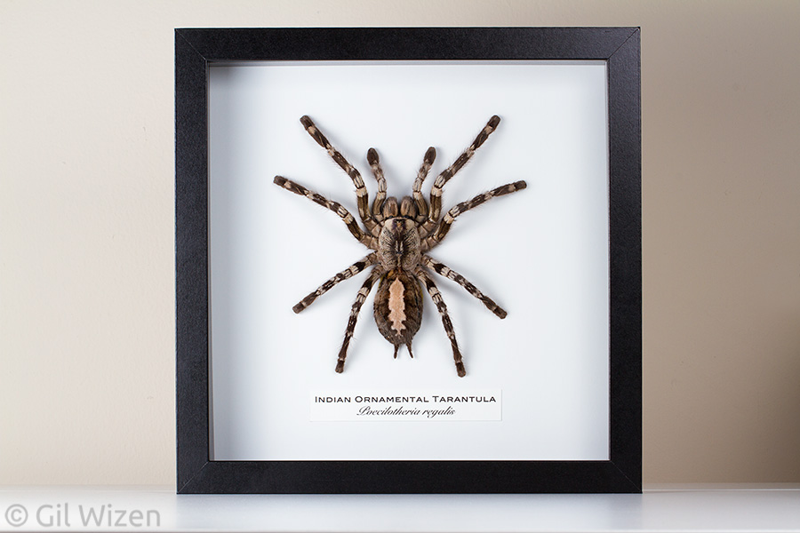
One of the framed tarantula molts I made this year. It was already sold before I got to finish it.

One of the framed tarantula molts I made this year. It was already sold before I got to finish it.

One thing that has definitely picked up pace is my shop page. I initially started that page in 2017 to see if there is any interest in my framed “Ethical Molts” (term coined by my dear friend Peggy Muddles, who also made the video below), and since then it has exploded. I have sold and shipped over 30 framed specimens, some went to distant locations overseas. Many times when I upload a new batch of frames it is sold within a few days. I cannot complain.


Final words

As you can see, I have been busy this year. Looking back, 2017 and most of 2016 were painful and unproductive years for me. 2018 marked a much-needed shift in the right direction. It is especially interesting to compare the summary posts from the two previous years to this one. Going back to my closing statement for 2017, a friend and I were discussing life goals and what counts as success. For many people, including my friend, financial prosperity counts as success. For me, success is not about getting rich. It is first about reaching stability (not necessarily financial), and then leaving something behind, making a positive impact on others. And in 2018, I felt I did exactly that. Thank you everyone for being a part of it. I am forever grateful for your support, and my life is so much richer because you are in it.

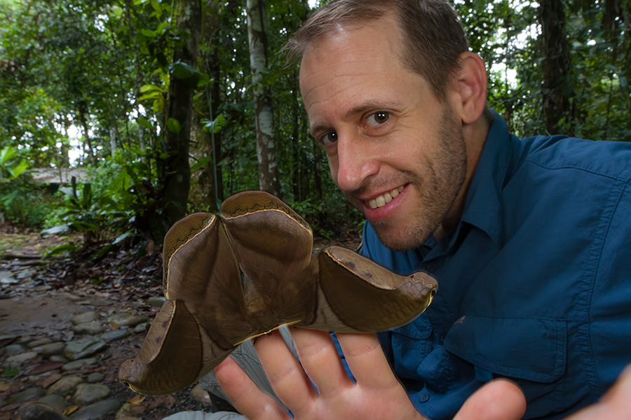
Me with a giant silk moth (Rhescyntis hippodamia) in Ecuador

Me with a giant silk moth (Rhescyntis hippodamia) in Ecuador

Little Transformers: Deinopis, the ogre-faced spider

Today’s Little Transformer is a little unusual. First off, it is a spider. This spider is so unique in its appearance and behavior that I am surprised it has not inspired any exaggerated depictions in popular culture. It spends most of its time hidden, posing as a harmless twig among the forest vegetation. It is so good at what it does, that unless it moves it would be very easily overlooked. However, when night falls this seemingly harmless twig transforms into a sophisticated killing machine. Meet Deinopis, the ogre-faced spider (also known as net-casting spider).

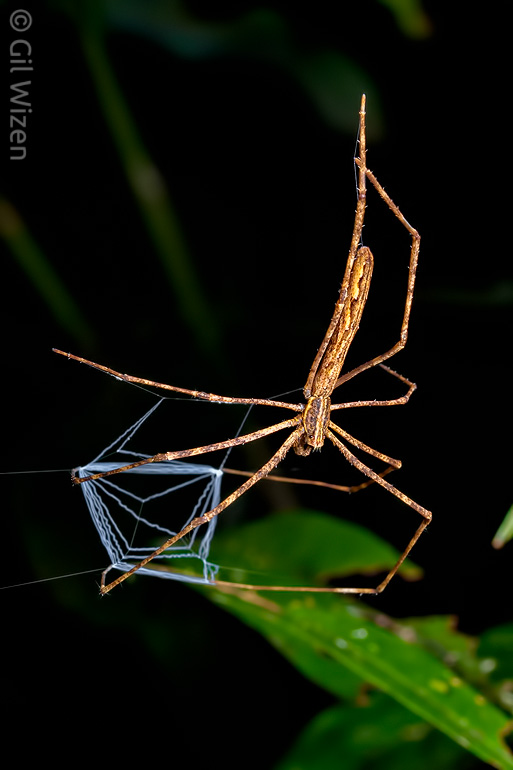
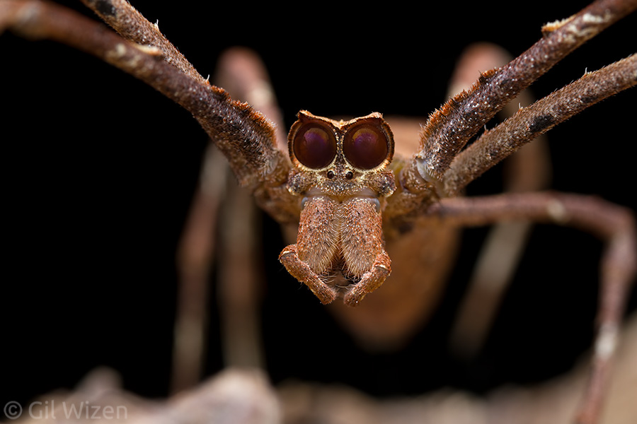
Net-casting spider (Deinopis sp.) from the Ecuadorian Amazon

Net-casting spider (Deinopis sp.) from the Ecuadorian Amazon

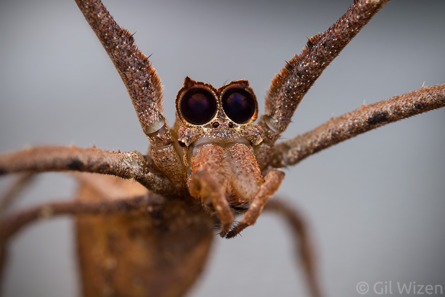
Net-casting spider (Deinopis spinosa) frontal view. Their eye arrangement is one of the weirdest of all spiders. Notice the lateral eyes are pointing down!

Net-casting spider (Deinopis spinosa) frontal view. Their eye arrangement is one of the weirdest of all spiders. Notice the lateral eyes are pointing down!

Ogre-faced spiders are found on every continent except Europe and Antarctica, but they occur mostly in warm regions of the southern hemisphere. Found primarily in Latin America, Africa, Madagascar, and Australia, these spiders all share the same appearance: brown color, elongated body with long forelegs, and an unmistakeable face. The small family Deinopidae contains only two genera: Deinopis, holding most of the species, and Menneus.
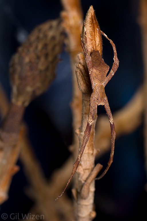
Net-casting spider (Deinopis spinosa) camouflaged as a twig or a dried leaf

Net-casting spider (Deinopis spinosa) camouflaged as a twig or a dried leaf

Interesting texture and patterns on the dorsal side of a net-casting spider (Deinopis spinosa). The legs are held tightly to form a typical 'X' shape at rest, making it look like the spider has only four legs.

Interesting texture and patterns on the dorsal side of a net-casting spider (Deinopis spinosa). The legs are held tightly to form a typical ‘X’ shape at rest, making it look like the spider has only four legs.

Deinopis are very unique among spiders for having superb vision, thanks to their huge median eyes.

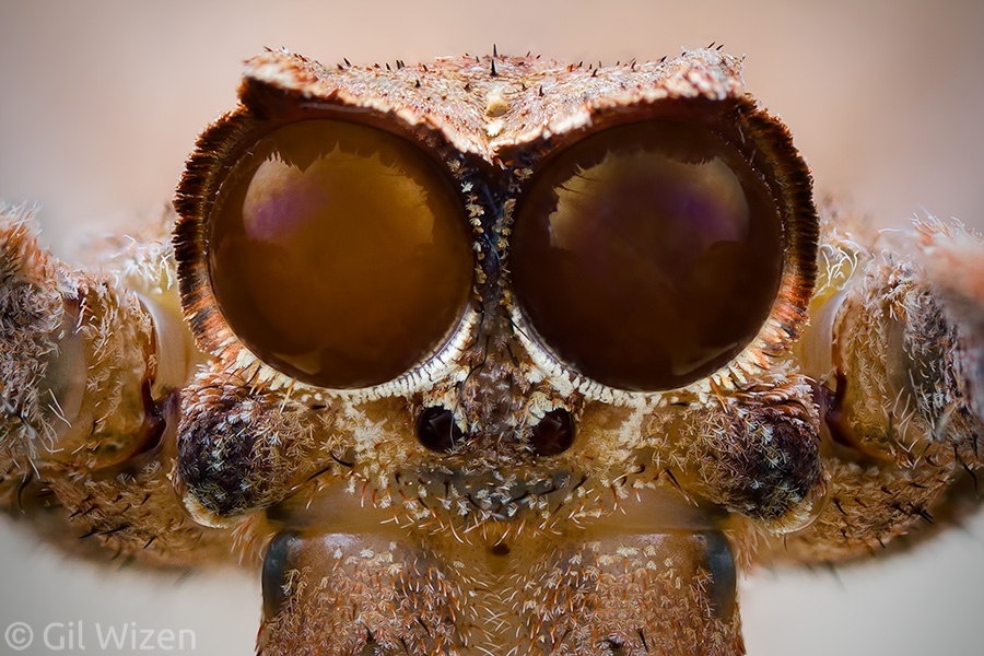
Net-casting spider (Deinopis spinosa). Those eyes... You can now understand why they are called ogre-faced spiders.

Net-casting spider (Deinopis spinosa). Those eyes… You can now understand why they are called ogre-faced spiders.

A closer look at the median eyes of a net-casting spider (Deinopis spinosa). Staring straight into your wretched soul.

A closer look at the median eyes of a net-casting spider (Deinopis spinosa). Staring straight into your wretched soul.

The big eyes are what gave these spiders their common name, and they are so big that it is easy to miss the other six eyes on the spider’s head. I did my best trying to capture the stone-cold expression on a Deinopis spinosa face, but also check out Michael Doe’s amazing work with the Australian species D. subrufa. These median eyes are extremely sensitive to light, despite lacking any reflective tissue behind the lenses. Instead, a light sensitive membrane is formed inside the eyes every night, and then gets broken down at dawn. This allows the spiders to track subtle movements in complete darkness during their activity hours, something that is essential for their unique hunting strategy. While somewhat close to other weavers, ogre-faced spiders do not construct a fixed web to trap their prey. Instead they make a rather small hand-net, a handkerchief if you wish, that they use to catch insects passing nearby. The silk constructing the net is not sticky but extremely fuzzy and flexible, thanks to a special comb-like structure on the spider’s legs that stretches and frizzle the silk as it is coming out of the spider’s spinnerets.

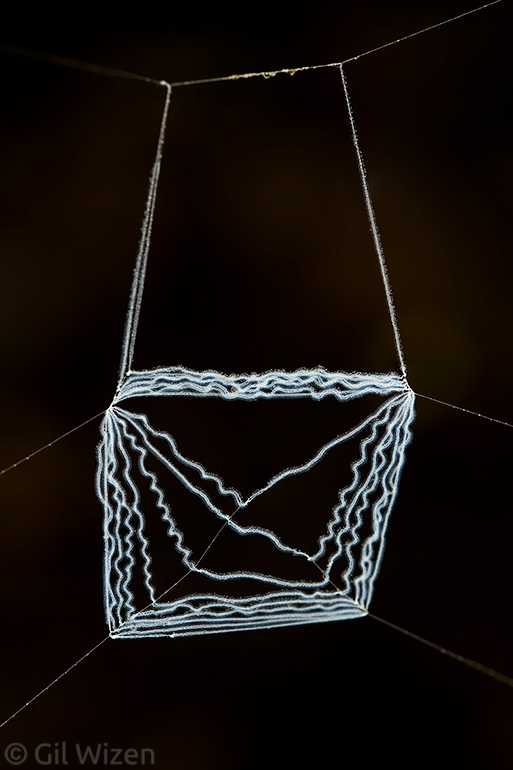
Net made by a net-casting spider for catching prey

Net made by a net-casting spider for catching prey

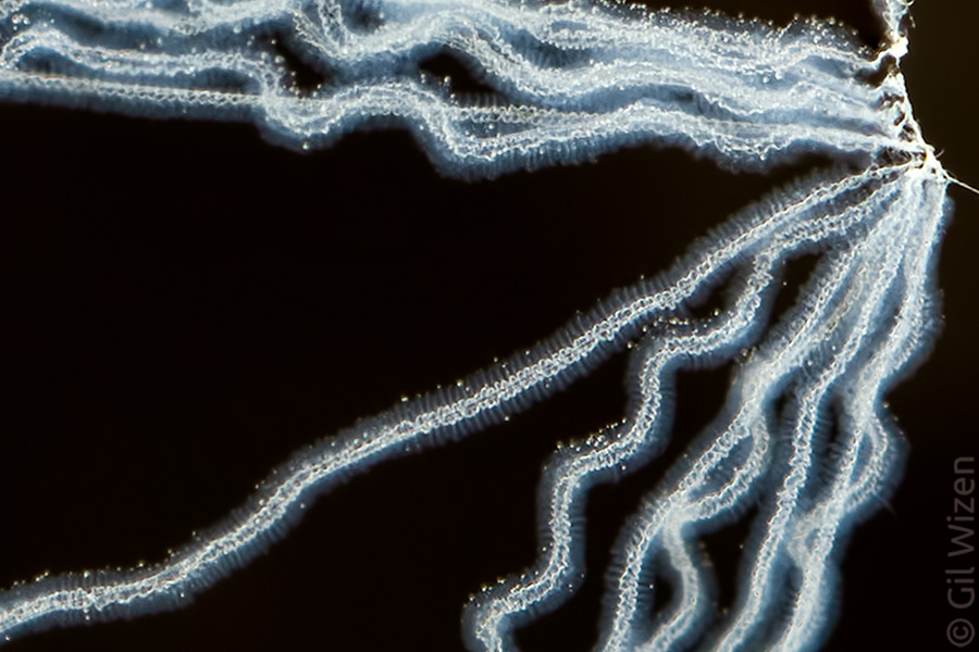
A closer look at the net reveals the woolly silk used to make it. If you look carefully you will notice that it is coiled like a spring, allowing the silk to be stretched and expanded to completely cover the prey.

A closer look at the net reveals the woolly silk used to make it. If you look carefully you will notice that it is coiled like a spring, allowing the silk to be stretched and expanded to completely cover the prey.

The spider usually shapes the net as a square, and holds it loose over a branch or a leaf where an insect is likely to walk.

Net-casting spider (Deinopis sp.) from Colombia

Net-casting spider (Deinopis sp.) from Colombia

Once it spots a suitable prey, the spider quickly stretches the net and snatches the passing insect by hand. The net can be stretched and expanded up to five times its original size without being torn, thanks to the special attributes of the silk. It entangles anything it touches. The spider is extremely fast in its response that sometimes it succeeds in capturing passing insects in mid-flight, again – completely by hand. You have to appreciate the speed and accuracy that goes into this hunting technique.

Net-casting spider (Deinopis sp.) ready for an insect to pass on a nearby branch. These spiders usually place themselves right above a possible walkway for arthropods. Photographed in Ecuador

Net-casting spider (Deinopis sp.) ready for an insect to pass on a nearby branch. These spiders usually place themselves right above a possible walkway for arthropods. Photographed in Ecuador

Net-casting spider (Deinopis sp.) from Honduras

Net-casting spider (Deinopis sp.) from Honduras

It is relatively difficult to witness this behaviour in the field, mainly because by observing at night we add another component to the equation – light. In fact, in all my trips to Latin America I have encountered these spiders many times, but only once I was able to see the spider hunting… and totally missing the prey insect. So you can imagine my excitement when I realized I was going to work with one of the species, Deinopis spinosa, while it is on display at the Royal Ontario Museum’s “Spiders: Fear & Fascination” exhibition. For several weeks I tried to get a glimpse of it feeding but without success. One day I decided to toss a cricket close to it before leaving the exhibit area and within a spilt second the spider responded and caught it! I was in awe. I had to find a way to record it on video for people to see. I enlisted Daniel Kwan, one of my colleagues at the museum who has more videography experience, and we set out to produce a short movie. It took us many attempts to get decent footage of the hunting behaviour. Many times the prey crickets tried to hide, and occasionally the spider would respond to them but miss. Even though feeding the spider in an artificial environment means we had more control, it was really difficult. It makes me wonder how long the spider must wait in the wild until it is able to catch a meal.

Also worth mentioning is genus Menneus from the same family. These spiders are much smaller than Deinopis and they lack the large median eyes, therefore they are not true ogre-faced spiders. However, they spin a catch net and use the same strategy for hunting prey. The genus contains only a handful of species, distributed mainly in Australia, but with some representation in Africa. Some of the species are quite beautifully patterned compared to the plain-looking Deinopis, and there are even green-colored species! You can find some photos of Menneus spiders at the bottom of this page.

Something I was thinking about while writing this post – why do I never encounter small deinopids in the field? It would be really cute if they had miniature nets for catching even smaller insects. Even when I look for information online and in the literature, it only concerns medium-sized juveniles and adults. Could it be that the small ogre-faced spiders actually have a different hunting strategy than that of larger individuals?

The Plot Thickens: This caterpillar ain’t big enough for the two of us

Some of my favorite insects to find while out in the field are hawkmoth caterpillars, or hornworms (named after the characteristic “tail”). They are big, squishy sausages that often show off dazzling colors, sometimes with interesting anti-predator adaptations like eyespots and mimicry. All these characters make the hawkmoth caterpillar look like a toy just waiting for you to play with. The sad truth is that being big and flashy in the natural world often comes with a price. There is danger lurking in every corner. Despite the bright colors and adaptations, birds and lizards do not hesitate to snatch the caterpillars from branches, pathogens and spores of entomophagous fungi scattered everywhere increase the chance for passive infections, and parasitoids are always on the lookout for chunky hosts for their offspring. And the reality is that many of the caterpillars we get to encounter outdoors are already infected with something. I learned this the hard way: as a kid I used to rear a lot of butterflies and moths collected as caterpillars in the field, and many times I was devastated to witness my cute pets being reduced into a sticky mess while wiggly worm-like creatures emerge from their bodies. Sometimes I wonder how lepidopterans manage to keep their populations stable with so many enemies around.

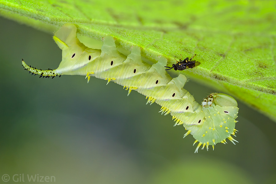
On one of my visits to the beautiful town of Mindo Ecuador, I came across a young hornworm. Despite finding it at daytime, the caterpillar remained calm (many hornworms do their best to disappear from plain sight during the day) so I decided to photograph it.

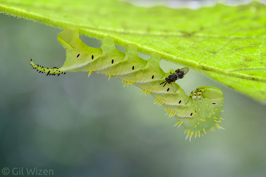
A cute hawkmoth caterpillar. See that black spot on the leaf? It is important to our story.

A cute hawkmoth caterpillar. See that black spot on the leaf? It is important to our story.

After taking a few shots I noticed a black splotch in the photo that I didn’t like, so I decided to change the angle of view. Little did I know this was a wasp that just arrived at the leaf to check out the caterpillar. A few photos later its identity became clear: It was a species of Brachymeria, a tiny wasp that belongs to the large parasitoid family Chalcididae.

The hawkmoth caterpillar being visited by a parasitoid chalcidid wasp (Brachymeria sp.)

The hawkmoth caterpillar being visited by a parasitoid chalcidid wasp (Brachymeria sp.)

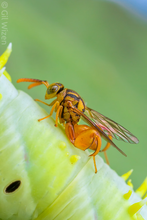
Chalcidid wasps can be easily recognized by their modified hindlegs that resemble mantids’ raptorial forelegs. The function of these structures is largely unclear. The adult wasps feed on nectar and other liquid foods, and do not use the legs for catching prey. There is a paper describing an interesting behavior in which the females use their legs in fighting over a host’s egg mass. Even more interesting are the last three paragraphs of the paper, with additional examples and hypotheses. It seems like there is no single function for these modified hindlegs and it really depends on the species and its biology. One example really stands out: “The female of Lasiochalcida igiliensis literally jumps into the jaws of antlions and holds the mandibles agape with her hind legs while ovipositing.”

Going back to our little Brachymeria and the hawkmoth caterpillar, at first the wasp just strolled peacefully on the leaf next to the caterpillar, but within a few minutes it hopped, quite literally, on the caterpillar and started walking on it, exploring its body surface while frantically moving its antennae.

The wasp jumped on the caterpillar's proleg and started crawling on its body

The wasp jumped on the caterpillar’s proleg and started crawling on its body

In general, the caterpillar doesn’t enjoy this attention, and often swiftly moves its head backwards in an attempt to drive the parasitoid away. It usually does not work. Once a caterpillar has been spotted and marked by a parasitoid as a host, it will be attacked (here’s a fantastic video showing this behavior, notice that the fly sitting nearby is another parasitoid of hornworms – a tachinid fly!).

A closeup of the parasitoid chalcidid wasp (Brachymeria sp.) as it was walking on the hawkmoth caterpillar

A closeup of the parasitoid chalcidid wasp (Brachymeria sp.) as it was walking on the hawkmoth caterpillar

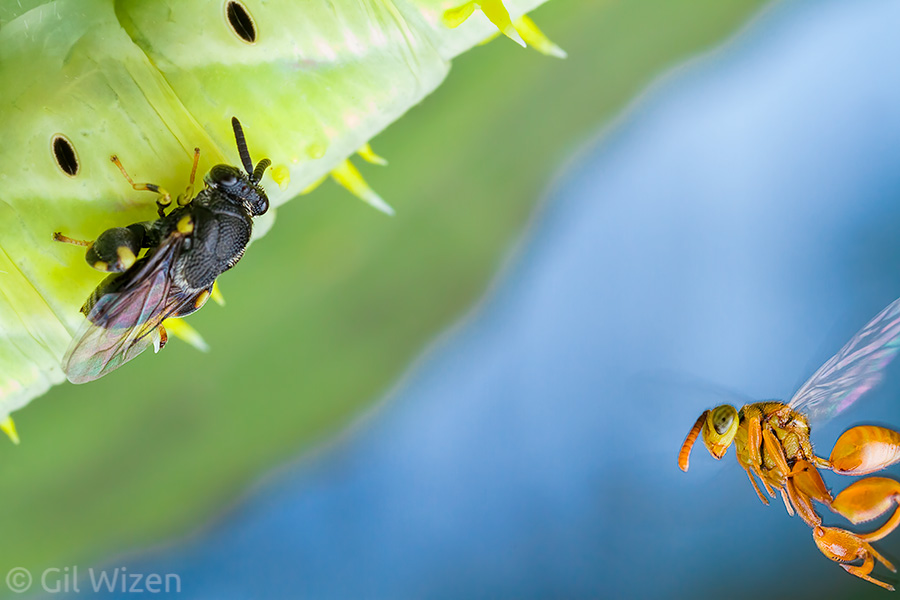
As I was taking photos of the tiny wasp antennating the caterpillar, from the corner of my eye I noticed a bright yellow object flashing in. A second wasp, a golden Conura species, swooshed into the scene and started harassing the busy Brachymeria wasp.

While the Brachymeria was busy exploring the caterpillar, another wasp (Conura sp.) rushed in to fight for it

While the Brachymeria was busy exploring the caterpillar, another wasp (Conura sp.) rushed in to fight for it

For a short while, the Conura striked from above repeatedly, yet the Brachymeria stood her ground. Eventually the Conura got fed up and attempted to grab onto the other wasp and pull her away from the host. After several tries she succeeded, and the two started swirling in the air, before the Brachymeria returned to her business on top of the caterpillar. The golden wasp did not give up and returned for a second attack and then a third.

The two chalcidid wasps (Brachymeria sp. and Conura sp.) fighting over the host. This was taken moments before the Conura grabbed the other wasp's head and dislodged it from the caterpillar.

The two chalcidid wasps (Brachymeria sp. and Conura sp.) fighting over the host. This was taken moments before the Conura grabbed the other wasp’s head and dislodged it from the caterpillar.

This was very exciting to watch, but to be honest I was waiting eagerly to see if the wasps would use their modified hindlegs during the fight. Unfortunately, I was not able to detect any special maneuvers that involved grabbing with those legs.

Why did this happen? There are several possible explanations. The simplest one is that there is a shortage of caterpillar hosts and the two wasps are competing for the same source of food for their larvae. However, an alternative explanation suggests that the caterpillar has already been infected with a parasitoid before the first wasp found it. Many chalcidid wasps are hyperparasitoids – they do not feed on the big hosts (the caterpillar) directly, but instead attack larvae of other parasitoids already feeding inside the host. In other words they are parasitoids of parasitoids.
Parasitoidception.
Watch this excellent video explaining the complex relationship between several wasp species living on a tobacco hornworm:

This can explain the intense antennation performed by the Brachymeria wasp on the caterpillar for a long period of time. Maybe the wasp was trying to determine if there are parasitoid larvae already present in there. One of the most common sights when it comes to infected hawkmoths is a caterpillar with a cluster of white silk cocoons dangling from its body. Those cocoons belong to braconid wasps, and there is a good chance that the Bracymeria wasp was after their larvae, as some species of in the genus are parasitoids of Braconidae. The golden Conura wasp could then compete for access to those parasitoid larvae or even go after the Brachymeria larvae. It can get pretty complicated with chalcidid wasps.

Hawkmoth caterpillar with cocoons of a braconid parasitoid wasp. The caterpillar is still alive, and can move its head to deter predators like ants and other parasitoids from approaching the developing wasps.

Hawkmoth caterpillar with cocoons of a braconid parasitoid wasp. The caterpillar is still alive, and can move its head to deter predators like ants and other parasitoids from approaching the developing wasps.

So who won in the end? The wasp that was more persistent. At the end of the fight the black Brachymeria wasp was nowhere to be seen, and the golden Conura wasp took over the caterpillar and started antennating it.

The winning chalcidid wasp (Conura sp.) with its hawkmoth caterpillar prize

The winning chalcidid wasp (Conura sp.) with its hawkmoth caterpillar prize

The interesting thing here is that members of genus Conura are usually associated with butterfly and moth’s pupae, yet the wasp here decided to chase off a competitor and take over a caterpillar.

Chalcidid wasp (Conura sp.) on a swallowtail butterfly pupa

Chalcidid wasp (Conura sp.) on a swallowtail butterfly pupa

Chalcidid wasp (Conura sp.) on a swallowtail butterfly pupa. This innocent face hides a dark secret.

Chalcidid wasp (Conura sp.) on a swallowtail butterfly pupa. This innocent face hides a dark secret.

Unfortunately, I had to leave the scene to catch a bus so I could not continue following this interaction. Without further observations, it is difficult to say with certainty what exactly was going on between the two wasps and the hawkmoth caterpillar. Parasitoids are so diverse, and many species have such complex biology. Even though several chalcidid wasp species are being studied closely as potential biological control agents, there are far more species out there about which we simply don’t know enough!


The Plot Thickens: Staring into the eyes of a dying Cephalotes

If you are an entomologist or an insect enthusiast, it is highly probable that you like ants. It is hard not to be impressed with their diversity, abundance, complex social structure and behaviors, as well as their interactions with other organisms. Ants are everywhere and do almost anything you can think of. To most people however, ants could not be any less exciting. They are often seen as a generic insect, with a relatively uniform appearance. They always show up when unwanted, find their way into our homes, take refuge in dark and hard to reach corners, and steal our food.
I like ants. I think they are fascinating creatures. But every now and then I find myself talking people into looking beyond “that boring-looking ant”, to try and catch a glimpse of their busy life. It is not always easy to communicate ants to the public (which is why I praise myrmecologists – people who study ants for a living), however I find that it is quite easy in the case of one ant genus in particular: Cephalotes.

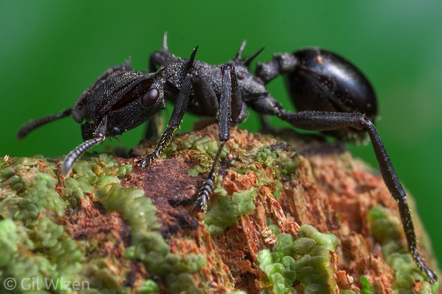
Turtle ant (Cephalotes atratus) from the Ecuadorian Amazon

Turtle ant (Cephalotes atratus) from the Ecuadorian Amazon

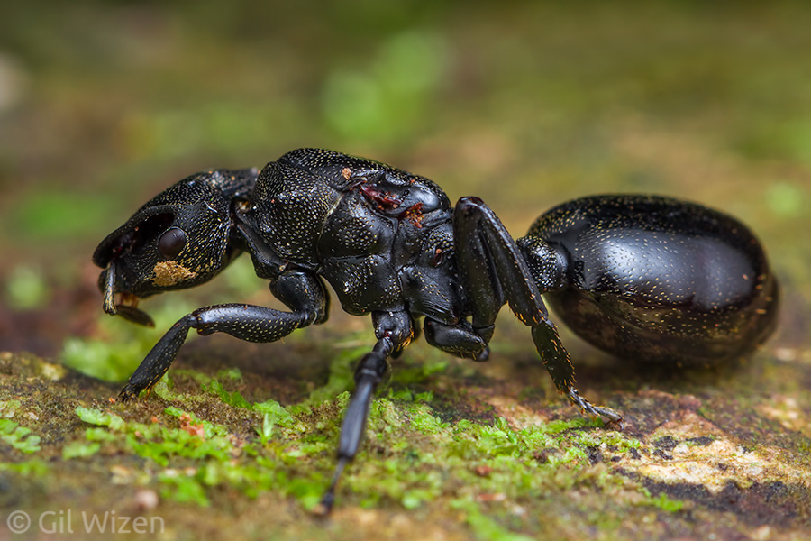
Cephalotes is a large genus of arboreal ants found in the neotropics. There are over 130 species, all inhabit tree hollows or utilize cavities in other plant tissues. Looking like they were designed by someone with overflowing imagination, they easily come off as cute. Their flattened head and armored body, often decorated with long sharp spines for protection, their thick legs and perfectly round abdomen, along with their matte color finish, give them the appearance of a plastic toy. In addition, Cephalotes ants move relatively slowly and cannot bite or sting, making them user-friendly. Can you ask for a more perfect ant?

The queen turtle ant (Cephalotes atratus) is bigger and bulkier than her workers. She also lacks the defensive spines.

The queen turtle ant (Cephalotes atratus) is bigger and bulkier than her workers. She also lacks the defensive spines.

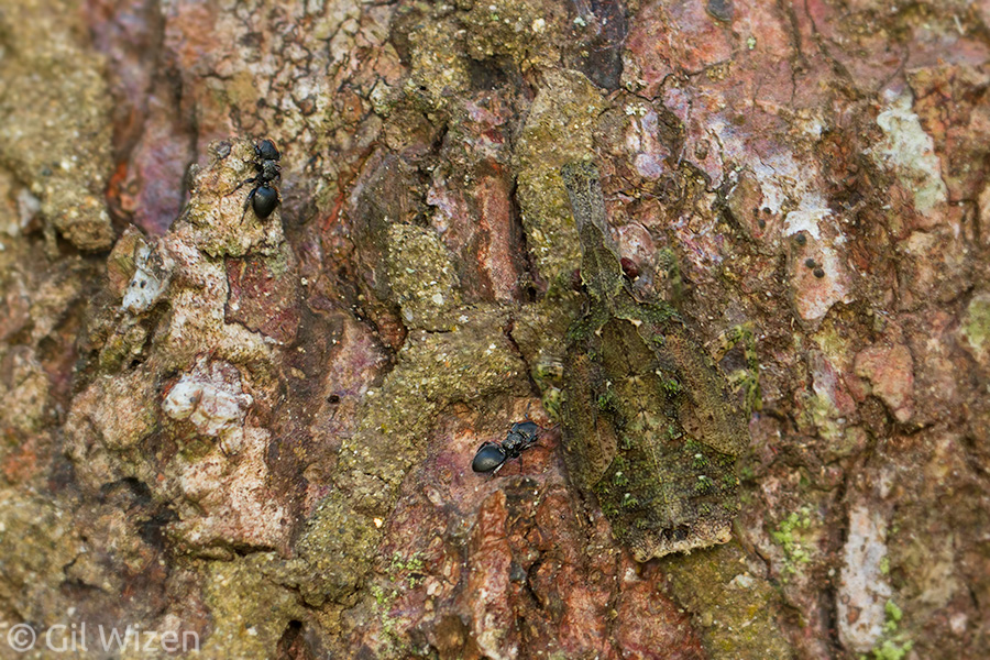
Turtle ant worker (Cephalotes atratus) foraging on a mossy tree trunk

Turtle ant worker (Cephalotes atratus) foraging on a mossy tree trunk

They are commonly known as turtle ants, but also got the name gliding ants, thanks to their incredible ability to parachute from high in the canopy and land back on the trunk of their home tree. Their unique body structure and flattened legs allow them to slow down and change their course while falling (some spiders can do the same, by the way). In some species the soldier cast evolved a large head to function as a living door, plugging the entrance to the nest.

Turtle ant soldier (Cephalotes sp.) from Colombia, showing a heavily armored body and a massive head

Turtle ant soldier (Cephalotes sp.) from Colombia, showing a heavily armored body and a massive head

The same turtle ant soldier (Cephalotes sp.) from the previous photo. These ants are built like tanks.

The same turtle ant soldier (Cephalotes sp.) from the previous photo. These ants are built like tanks.

In regards to interspecific interactions, Cephalotes ants are often seen tending sap-sucking hemipterans such as membracids and small fulgorids to gain access to sugary secretions from those insects. They also act as the model in a mimicry complex, where crab spiders masquerade as the ants in order to sneak up and prey on them.

Cute Cephalotes workers visiting a camouflaged fulgorid planthopper nymph

Cute Cephalotes workers visiting a camouflaged fulgorid planthopper nymph

Portrait of a turtle ant (Cephalotes atratus). How can you not fall in love with them?

Portrait of a turtle ant (Cephalotes atratus). How can you not fall in love with them?

Did I mention they are cute? I have written before that you should never become too attached to insects you encounter in the field. And as much as I love the adorable Cephalotes ants, it is important to remember that there are many dangers lurking for them in the forest. During my recent trip in Colombia, I stumbled upon a Cephalotes nest in a tree outside my room. The ants were very active and did not present good photographic opportunities.

Turtle ant (Cephalotes sp.) from Colombia. How adorable!

Turtle ant (Cephalotes sp.) from Colombia. How adorable!

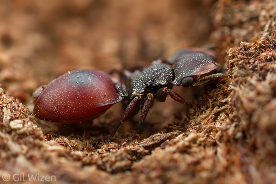
One of them however, stood out among the rest. There was something different about its behavior. This worker moved franticly in what appeared to be an aimless run. It did not follow the other workers, and seemed more interested in reaching a higher spot on the tree. I collected the ant for a closer look, and once I inspected her carefully I believe I found the culprit for her unusual behavior. This ant had a reddish abdomen, as opposed to the black abdomen of her sisters. The red color, coupled with erratic behavior suggests this worker has been infected with a parasite, a nematode worm.

Turtle ant (Cephalotes sp.) infected with a parasitic nematode worm, showing a swollen red abdomen. Compare to the healthy worker in the previous photo.

Turtle ant (Cephalotes sp.) infected with a parasitic nematode worm, showing a swollen red abdomen. Compare to the healthy worker in the previous photo.

The parasitic worm lives and breeds inside the body of birds, which spread the worm’s eggs in their droppings. The ants collect nutrients from the bird droppings (along with the eggs) and feed them to their larvae, where the worm matures. In order to complete its life cycle the parasite needs to return into a bird’s body, so it changes the host ant’s appearance to look like a ripe red fruit, and causes it to climb higher on the tree to become more accessible to hungry birds. As much unique character this worker ant might have had, the sad truth is that it was destined to die prematurely. And there was nothing I could do about it. There is a great lesson here – sometimes, the raw essence of nature is difficult to take in. We would like to see it as a peaceful place where all the animals and plants live together in harmony. But the reality is that nature is harsh. It is full of conflict, violence, disease, and death. And we must accept it as an integral part of the world we live in.

Cephalotes ants offer a great opportunity to peek into the life of a small insect and learn about its survival (as well as failure) in various habitats. Before I end this post, there is one thing I would like clarified – going back to their name, why did Cephalotes get the name turtle ant, whereas some leaf beetles were named tortoise beetles? Is there any justification for the turtle designation when it comes to the ants? After all, both insects are terrestrial. If there is an etymologist in the audience, maybe you can help the entomologist?

Little Transformers: Forcipomyia, the midge that turns into a balloon

It is time to introduce another Little Transformer! I know what you are thinking. Am I ever going to run out material for these blog posts? Maybe. Probably not. As long as there are arthropods around, their life history and morphological diversity guarantees that I will always find examples for interesting deceptions and transformations. Up until now I mostly focused on animals that can change form quickly, assuming the appearance of something else as a defense response against predators and to avoid detection. The case presented in this post is a little different because it does not follow a quick change of form, but rather a slow one, over the course of a life stage. I should be cautious here, because under this definition every insect that goes through complete metamorphosis from larva to adult can be considered a Little Transformer (butterflies, beetles etc’). Even amphibians fall under this loose definition. And to some extent they ARE transformers, because the changes they go through during development are extreme. But this is not the topic for this series of posts. When I talk about a big change happening within a life stage, I mean that the animal starts as one thing, and by the end of the stage its appearance and function has changed into something else completely. And no example is better to show this than the parasitic midges of the genus Forcipomyia.

Biting midge (Forcipomyia sp.) feeding on the hemolymph of a moth caterpillar. Photographed in Belize

Biting midge (Forcipomyia sp.) feeding on the hemolymph of a moth caterpillar. Photographed in Belize

Here is the Forcipomyia midge with the whole caterpillar to give a better sense of scale

Here is the Forcipomyia midge with the whole caterpillar to give a better sense of scale

Forcipomyia is a large genus in the midge family Ceratopogonidae, with a worldwide distribution and diverse habitat preferences. There are now over 1,000 described species of Forcipomyia. The adults of some species are known as important pollinators of cacao and other plants of economic importance in tropical and subtropical areas. However, many species in the genus are blood-feeders, somewhat characteristic to ceratopogonids as the common name to the family suggests (biting midges). These parasites have interesting relationships with different insect hosts, and they can be found feeding on the hemolymph (insect blood) of grasshoppers, katydids, stick insects, butterflies, true bugs, and even skittish dragonflies. In fact, these interactions are so fascinating and overlooked, that only after spending some time in the field one can notice the midges have a preference for certain host species to feed from.

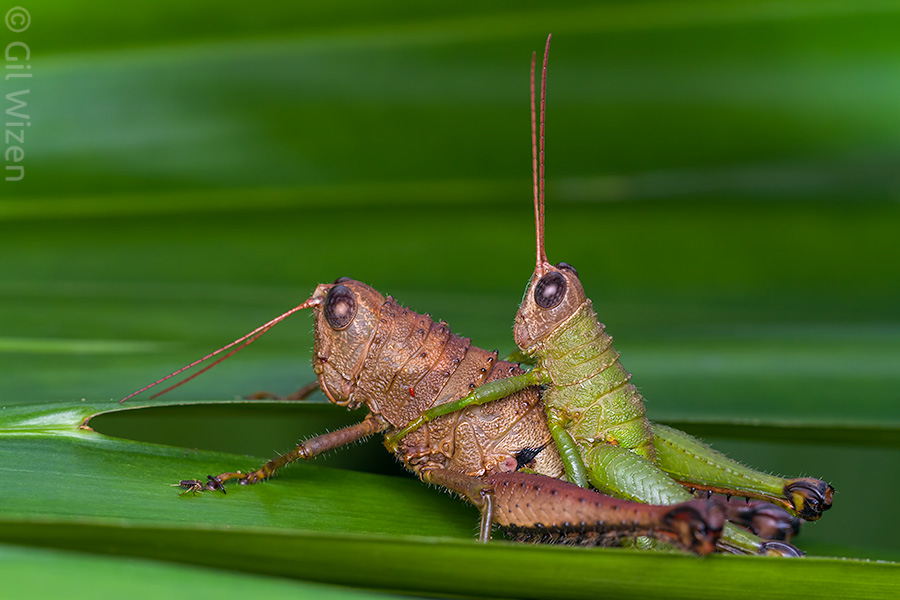
Sometimes the biting midges sneak into the photo without me noticing. I photographed these mating grasshoppers (Cloephoracris festae), but they have an accompanying Forcipomyia. Can you spot it?

Sometimes the biting midges sneak into the photo without me noticing. I photographed these mating grasshoppers (Cloephoracris festae), but they have an accompanying Forcipomyia. Can you spot it?

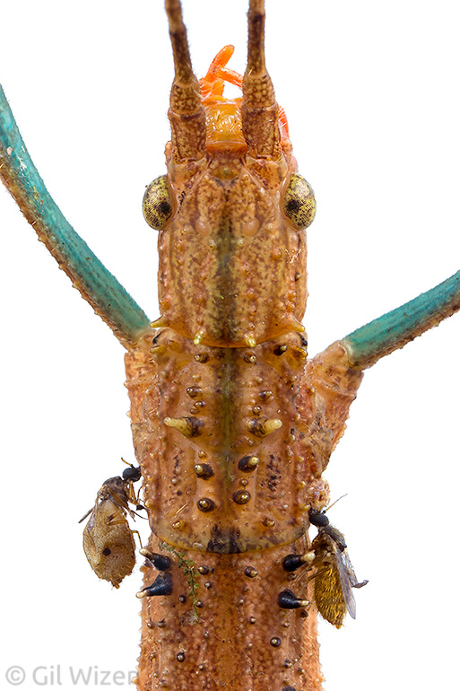
But let’s go back to the transformation they go through, because in one group of species, subgenus Microhelea, it is truly remarkable. The female Forcipomyia midge begins her adult stage with an active lifestyle. She flies about in the forest, feeding on nectar from small flowers. As days go by, she starts craving for blood and search for insects to bite. When she locates her preferred host, using her serrated mouthparts she proceeds to bite it in an area that has soft tissue: antennae, legs joints, wing veins, or between body segments. Once she found the right spot that will fulfill her dietary needs, the female midge attaches to it firmly, and… doesn’t let go, thanks to specialized claws on her feet. She sucks and gulps the insect’s blood, filtering the nutrients and secreting the excess fluids as clear droplets.

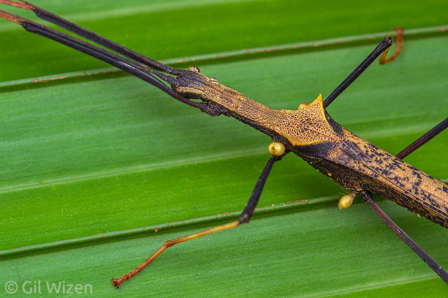
Tick fly (Forcipomyia sp.) feeding on the hemolymph of a walking stick

Tick fly (Forcipomyia sp.) feeding on the hemolymph of a walking stick

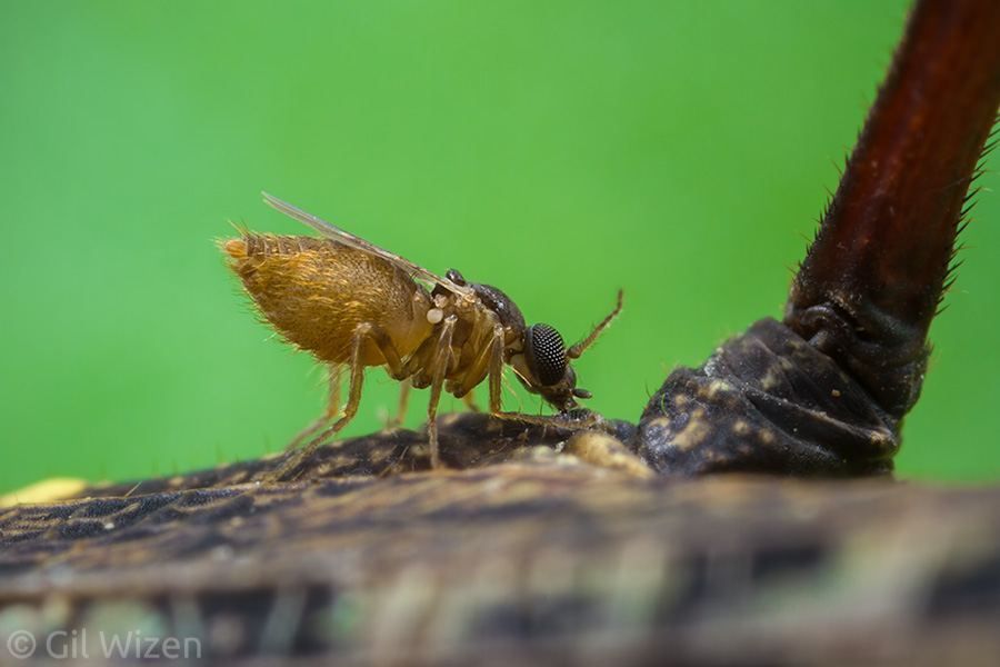
The midge stays attached like this for quite a while, and soon this sessile lifestyle starts taking its toll on the small parasite. She starts to put on weight. Then, she usually loses her wings – she will not need them anymore because the added mass from the developing eggs prevents her from taking off.

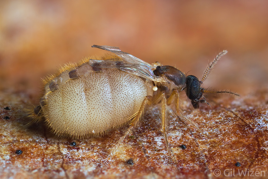
Female Forcipomyia swelling while feeding. She lost her wings but can still use her legs to hold firmly onto the host

Female Forcipomyia swelling while feeding. She lost her wings but can still use her legs to hold firmly onto the host

Forcipomyia getting fatter... but not quite there yet

Forcipomyia getting fatter… but not quite there yet

As she continues to swell like a grapefruit, the Forcipomyia midge also losses the ability to use her legs. She does not need to leave anyway, but she is so bloated that she cannot even hold onto the body of the host, and the only thing keeping the two connected are the midge’s mouthparts.

Female tick fly (Forcipomyia sp.) at the final stage of feeding. Her legs released their grip on the host and at this point the midge has fully transformed into a passive parasite that looks like a balloon.

Female tick fly (Forcipomyia sp.) at the final stage of feeding. Her legs released their grip on the host and at this point the midge has fully transformed into a passive parasite that looks like a balloon.

Stick insect (Pseudophasma bispinosum) carrying tick flies (Forcipomyia sp.) at different stages of feeding. Photographed in Ecuador

Stick insect (Pseudophasma bispinosum) carrying tick flies (Forcipomyia sp.) at different stages of feeding. Photographed in Ecuador

At this point, the engorged biting midge is no different than a tick, and indeed many refer to these parasitic Forcipomyia as tick-flies. Sometimes I like to imagine these fat dipterans disconnecting from their host and floating upwards like a balloon filled with helium, reaching above the forest canopy and flying into space. In reality, the exact opposite happens. The Forcipomyia female eventually leaves the host and drops to the ground, where she lays her eggs and finishes her role. And the male Forcipomyia? They are mostly unknown. Because males are never found feeding on insect hosts, it is safe to assume that they do not feed on blood, and prefer to keep a vegan diet of sweet nectar.

An engorged female tick fly (Forcipomyia sp.) after dropping from its host

An engorged female tick fly (Forcipomyia sp.) after dropping from its host

Tick fly (Forcipomyia sp.) engorged with hemolymph viewed from above

Tick fly (Forcipomyia sp.) engorged with hemolymph viewed from above

What about the larvae, are they parasites too? The majority of the research on biting midges has focused on the adults, due to their economic and medical significance, as well as their important role in aquatic ecosystems. Larvae of most ceratopogonids are unknown because finding them in their natural habitats can be challenging. They usually inhabit aquatic and semiaquatic habitats, but in the case of Forcipomyia the larvae are terrestrial and prefer to feed on moist detritus and organic matter under bark or in moss. In some species they feed on algae.

This stick insect is staring at me with tired eyes. I wonder if it is aware of the two hitchhikers it is carrying?

This stick insect is staring at me with tired eyes. I wonder if it is aware of the two hitchhikers it is carrying?

With so many aspects of their life history still unknown, and especially due to their ecological and economical importance, you would expect to see more active research on Forcipomyia. The bad news is that there is not enough research going on. A few years ago, I approached Dr. Stephen Marshall, a dipterologist from University of Guelph, and suggested doing a PhD study about Forcipomyia’s biology, phylogenetics, and their relationships with their hosts. I was politely refused, unfortunately. I still believe there is potential for a cool project involving Forcipomyia, maybe someone will pursue it in the future.

Little Transformers: Lamprosoma, the living Christmas ornament

Ah, the joy of transforming beetles. The first Little Transformer that opened this series of posts was a beetle – a Ceratocanthinae pill scarab that transforms into a perfect sphere and drops off to escape predators. It is an impressive evolutionary achievement that merges a successful body design and anti-predator behavior. I should mention though that many beetle species from other families use this strategy to avoid predation, some more successfully than others. One such example is a genus of small beetles from the leaf beetle family (Chrysomelidae): Lamprosoma.

Shiny leaf beetle (Lamprosoma sp.) from the Ecuadorian Amazon

Shiny leaf beetle (Lamprosoma sp.) from the Ecuadorian Amazon

When I first encountered a Lamprosoma beetle I thought it was a piece of plastic that someone discarded in the rainforest. There is something almost artificial about their appearance, shiny metallic colors combined with a compact shape. Not all species are colorful, by the way. The genus contains about 130 species, all with a neotropical distribution, some of which are completely black in color. With a body length of less than 1cm they are easy to miss in the dense vegetation of the tropical forest. Nevertheless, over the years I have encountered them more and more frequently. Unfortunately for me, identifying these beetles to the species level requires an expertise that I do not have, because there are many similar-looking species, and possibly also new species that have not been described yet.
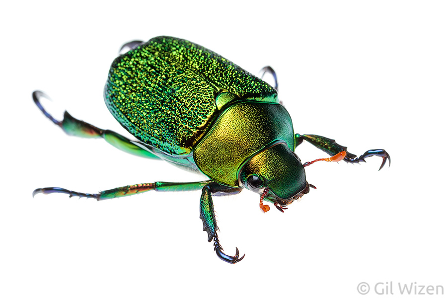
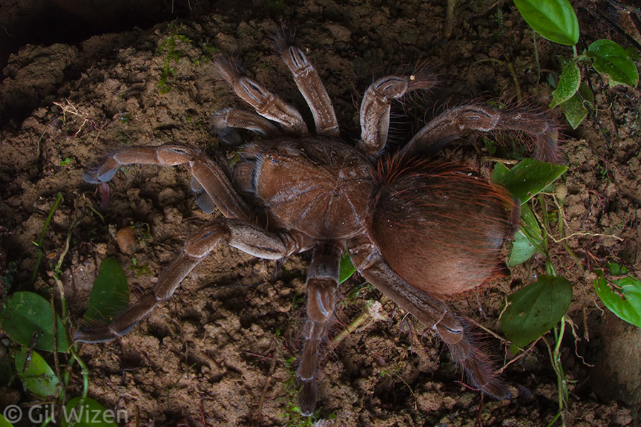
Shiny leaf beetle (Lamprosoma sp.) from Honduras

Shiny leaf beetle (Lamprosoma sp.) from Honduras

The beetles are dome-shaped, and have very short legs. I think “cute” is the best way to describe them. As mentioned above, Lamprosoma can transform into a ball when threatened. In contrast to Ceratocanthinae beetles that have dedicated grooves to hold the legs and head in place, members of genus Lamprosoma have no such features. The beetle tucks in its head and holds its legs tightly close to its body, making it a neat impenetrable package.

Shiny leaf beetle (Lamprosoma sp.), a ventral view showing how neatly they press their legs against the body when forming the ball

Shiny leaf beetle (Lamprosoma sp.), a ventral view showing how neatly they press their legs against the body when forming the ball

Shiny leaf beetle (Lamprosoma sp.) in ball-mode. Mimicking a Christmas ornament.

Shiny leaf beetle (Lamprosoma sp.) in ball-mode. Mimicking a Christmas ornament.

In species with shiny metallic colors it is hard not to see the resemblance to the glass balls used as Christmas ornaments (maybe an idea for a future product?). Once the danger is out of sight, the beetle loosens its legs and walks away.

Shiny leaf beetle (Lamprosoma sp.) transformation sequence from ball-mode to beetle-mode. How can you not fall in love with those stubby feet?

Shiny leaf beetle (Lamprosoma sp.) transformation sequence from ball-mode to beetle-mode. How can you not fall in love with those stubby feet?

Lamprosoma are phytophagous beetles, meaning that they feed on plants. Both adults and larvae feed on leaves, and can be potential pests due to damage they can cause to foliage. The species shown here seem to be associated with cacao trees, and were found under leaves during the day. While the adults are very showy, the larvae are cryptic to avoid predators: they construct a case from frass and wood debris, and carry it around throughout their lifetime. The case is often shaped like a bent thorn, and blends perfectly with the branches the larvae live on. When threatened the larva retreat into the case and hold it firmly against the branch, preventing predators (such as ants and wasps) from accessing inside.

Another example of Lamprosoma sp. in ball-mode

Another example of Lamprosoma sp. in ball-mode

Shiny leaf beetle (Lamprosoma sp.). Full beetle-mode!

Shiny leaf beetle (Lamprosoma sp.). Full beetle-mode!